
Albendazole
General Information about Albendazole
Aside from treating tapeworm infections, Albendazole has additionally been used within the treatment of different parasitic infections, corresponding to roundworm, hookworm, and whipworm infections. It has also been used to deal with different conditions like cysticercosis (an an infection attributable to tapeworm larvae) and neurocysticercosis (a extreme form of cysticercosis affecting the central nervous system).
Albendazole is generally protected and well-tolerated, but like several medicine, it could trigger unwanted effects in some people. Some of the widespread side effects embrace nausea, vomiting, abdominal ache, headache, dizziness, and diarrhea. More serious side effects similar to fever, chills, and allergic reactions are uncommon however can happen. It is crucial to speak with a healthcare skilled if these or any other unwanted effects are skilled through the course of remedy.
Overall, Albendazole has been proven to be a extremely effective treatment within the treatment of tapeworm infections. Its capability to target and get rid of different varieties of tapeworms makes it a most popular alternative for healthcare professionals globally. However, prevention is all the time higher than treatment, and to avoid tapeworm infections, it is important to totally cook meat and keep proper hygiene practices. If you think that you might have a tapeworm an infection, search medical attention and observe the prescribed therapy routine to ensure a whole restoration.
One of probably the most generally used medications for treating tapeworm infections is Albendazole. This drug belongs to a category of medicines called anthelmintics, which work by killing the tapeworms or stopping them from growing and reproducing. Albendazole is particularly effective towards a broad variety of tapeworms, together with Taenia saginata (beef tapeworm) and Taenia solium (pork tapeworm), among others.
Albendazole is a widely recognized and generally used medicine in the remedy of parasitic infections caused by tapeworms. This medication has been proven to be highly efficient in eliminating tapeworm infections, which could be notably troublesome and detrimental to a person's well being.
Tapeworm infections, also called cestodiasis, are caused by a sort of worm that may develop and reside in a human's intestines. These worms can vary in size from a couple of millimeters to several meters in size and are transmitted through consuming undercooked or contaminated meat, notably pork and beef. Once inside the physique, tapeworms can survive and reproduce for years, causing a spread of unpleasant symptoms.
This medicine is available in tablet kind and is normally taken orally with meals. The dosage and duration of treatment could range depending on the sort of tapeworm infection and the severity of the symptoms. It is necessary to follow the prescribed regimen carefully to make sure the effectiveness of the treatment.
Albendazole works by inhibiting the formation of microtubules, essential buildings that tapeworms want to take care of their form and move around in the body. This prevents the worms from absorbing glucose, which in the end leads to their death. The medicine can also be thought to have an immunosuppressive impact, which helps to reduce back the inflammation and harm attributable to the tapeworms in the body.
These conditions should be considered in evaluating each patient presenting with these deformities www.hiv infection symptoms albendazole 400 mg purchase with visa, particularly if the problem is unilateral. This phenomenon is accentuated by overlying callosities that develop as a result of abnormal weight bearing. Neurologic examination may reveal motor weakness, most often involving the anterior tibial, toe extensor, and peroneal muscles. Logical treatment necessitates identifying and treating the underlying pathologic condition when possible. Nonsurgical measures for managing the deformities and ameliorating the symptoms consist of the wearing of customized shoes and use of a metatarsal bar to relieve pressure on the metatarsal heads and to correct the extension deformities at the base of the toes. The contralateral foot exhibits a metatarsus adductus deformity, giving the feet a "windswept" appearance. Accessory Tarsal Navicular An accessory tarsal navicular results from formation of a separate ossification center on the medial aspect of the developing tarsal navicular at the insertion site of the posterior tibial tendon. The condition is not uncommon and is usually associated with a pes planus deformity. Clinically, patients exhibit a bony prominence on the medial aspect of the foot that tends to rub on the shoe, thus producing a painful bursa. Radiographs reveal either a separate ossification center or bone medial to the parent navicular, or a medial projection of the navicular when fusion has occurred. Long-term improvement can be obtained by wearing soft, supportive shoes with longitudinal arches and a medial heel wedge. Those relating to genetic, endocrine, collagen vascular, neurologic, and hematologic problems are discussed in their respective chapters. A, Laxity of the soft tissue structures of the foot results in a loss of the normal longitudinal arch and pronation or eversion of the forefoot. B, Viewed from behind, the characteristic eversion of the heels is appreciated more readily. A, A bony prominence produced by the formation of a separate ossification center of the tarsal navicular is present over the medial aspect of the midfoot. B and C, Anteroposterior and lateral radiographs of the foot demonstrate the accessory navicular. The posterior tibialis tendon attaches to the small accessory bone and may contribute to continued irritability and tenderness in this area. Despite the more than 300 types of identified skeletal dysplasias, many individuals with a presumed skeletal dysplasia remain unclassified. Although skeletal dysplasias comprise a heterogeneous group of disorders, two major categories exist: osteochondrodysplasia and dysostosis. The osteochondrodysplasias result from abnormal growth and development of bone and/or cartilage. These are progressive and generalized disorders and are the focus of this section. There are 33 groups of osteochondrodysplasia and three categories of dysostosis in the current classification system. Although individual skeletal dysplasias are rare, as a group they are relatively common. The incidence of all skeletal dysplasia is approximately 1 case per 4000 to 5000 births. A Danish study found that skeletal dysplasias represented 9% of the Danish population and that the incidence of congenital generalized skeletal dysplasias at birth was found to be 75. Because a proportion of skeletal dysplasias are lethal, the prevalence in the general population in that study was found to be much lower (33 per 100,000 population). Achondroplasia is usually regarded as the most common nonlethal skeletal dysplasia worldwide. Diagnosis Accurate diagnosis can be important for genetic counseling regarding future pregnancies and is helpful in predicting the clinical course, as well as in aiding in treatment strategies for complications. Diagnosis of specific skeletal dysplasias can be challenging because of limited availability of genetic testing. Often diagnoses are made on the basis of distinctive radiographic and physical findings. In addition, when growth halts after puberty, it is difficult to distinguish radiographically between the types of skeletal dysplasias, making it important to make a diagnosis as early as possible. Because diagnosis often relies on radiographic findings, it is important to obtain a skeletal survey of any infant or child in whom a dysplasia is suspected. Prenatal detection of a skeletal dysplasia is important because it determines the obstetric and perinatal management of an affected fetus. Because up to 30% of skeletal dysplasias can be lethal, accurate diagnosis is imperative for decision-making regarding possible termination. Unfortunately, prenatal diagnosis of specific skeletal dysplasias can be even more challenging than postnatal diagnosis. The deformity is often a feature of neuromuscular disorders, as in this case where it is the result of Charcot-Marie-Tooth disease. B, In addition to the high arches and varus (inverted) heels seen in the view of the plantar surface, the prominence of the metatarsal head region is apparent. Callosities have developed over the lateral borders of the feet as a result of abnormal weight bearing. Prenatal ultrasound of a suspected skeletal dysplasia involves systematic imaging of the long bones, thorax, hands and feet, skull, spine, and pelvis. Evaluation of thoracic dimensions revealing a hypoplastic thorax is suggestive of severe or lethal skeletal dysplasias. This leads to pulmonary hypoplasia and is a frequent cause of death in patients within the first year of life. The mainstay of prenatal diagnosis remains two-dimensional ultrasound, but it has a sensitivity of only 60%.
Mild to moderate isolated hydronephrosis is defined by an anteroposterior diameter between 5 to 15 mm without calyceal dilation antiviral cream for genital herpes 400 mg albendazole sale, normal renal cortical thickness, and no associated abnormal renal echotexture, hydroureter, or thickened bladder wall on ultrasonography at 1 week after birth. Typically, 94% of such cases resolve spontaneously during the first 12 to 14 months of life. PotterSequence Potter sequence results from fetal compression secondary to decreased fetal urine production (oligohydramnios). In the extreme example of bilateral renal agenesis the virtual absence of amniotic fluid during fetal life leads to Potter syndrome. This infant with bilateral multicystic dysplasia died at 12 hours of age with pulmonary insufficiency. The altered facies produced by the fetal compression syndrome of oligohydramnios includes small, posteriorly rotated ears, micrognathia, a beaked nose, and wide-set eyes. Hereditary and Metabolic Disorders CysticKidneyDisease Five of the most common and most important forms of renal cystic disorders are presented in this section. Cystic renal dysplasia has no well-defined inheritance pattern and is often associated with other syndromes. Renal cystic disease may also be an important component of many syndromes (Table 14. The renal cysts are quite small and numerous and often result in massive renal enlargement and palpable flank or abdominal masses compressing the abdominal contents and diaphragms. The majority of cases are severe and are often detected by antenatal ultrasonography after 24 weeks of gestation. These newborns may present with oligohydramnios or Potter-like phenotype (see the earlier section), including anuria or oliguria, and pulmonary hypoplasia. Less severe cases can present with abdominal distention due to renal enlargement and mainly tubular dysfunction. Hyponatremia often occurs in infancy and may be related to nonosmotic release of vasopressin, particularly in the setting of pulmonary disease, excessive renal salt wasting, and extracellular volume contraction. Kidney is enlarged with increased echogenicity with loss of corticomedullary differentiation. It is a leading cause of renal failure in the United States and affects 13 million people worldwide. Although most cases are diagnosed in adulthood, symptoms are increasingly recognized in early childhood. Within families, there is much variability in the phenotypic expression of clinical course, severity of renal disease, and extrarenal manifestations. With current high-resolution renal ultrasonography, up to 90% of affected individuals younger than 20 years old have detectable cysts. Pathologically, the cysts become very large and asymmetrical and involve all parts of the nephron and collecting duct. Similarly rare are extrarenal manifestations, including rupture of intracranial aneurysm, colonic diverticula, and symptomatic mitral valve prolapse. Multiple cysts of various size are seen throughout the cortex and medullary regions. CysticRenalDysplasia Abnormal renal morphogenesis can lead to absent parenchyma (aplasia), deficient parenchyma (hypoplasia). Thus, despite the presence of cysts, kidney sizes may be too small to appreciate by bimanual examination. When bilateral, these renal disorders may present early in life with poor weight gain, pallor, emesis, and tachypnea resulting from metabolic acidosis. Urine output is often maintained or increased due to concentrating defects, but correlates poorly with renal function reflected by the serum creatinine level. Renal hypoplasia is often an isolated disorder without other systemic or genitourinary tract anomalies, whereas obstruction of the gastrointestinal or genitourinary tract occurs frequently in children with renal dysplasia. Less common anomalies may include trisomy 21, tracheoesophageal fistula, ventricular septal defect, and lumbosacral dystrophies. Cystic dysplasia may be a major component of several syndromes with distinct additional malformations (see Table 14. The overall risk for siblings of children with isolated forms of dysplasia or hypoplasia is usually less than 10% but is generally higher than the general population if one of the parents has renal aplasia or other structural kidney disease. MulticysticDysplasia Multicystic dysplasia, usually unilateral, is the most common cystic disorder in children, as well as the most common cause of abdominal mass in newborns. Multicystic dysplasia represents an extreme case of renal cystic dysplasia in which there is complete loss of the normal renal architecture with microscopic primitive ducts, fibrosis, islands of ectopic cartilage, and usually ureteral atresia. This condition is most commonly discovered by prenatal ultrasonography, or it may be diagnosed during the neonatal period after palpation of a "lumpy" intraabdominal mass of variable size that often transilluminates. The parenchyma in the upper pole is normal, but microscopic examination of the lower pole showed several morphologic features of dysplasia. Very large multicystic kidneys can interfere with respiration or produce mechanical intestinal compression. Radionuclide scanning and renal ultrasonography are usually sufficient to establish the diagnosis. The unaffected contralateral kidney is usually hypertrophied and has normal corticomedullary differentiation and no evidence of obstruction. Obstructive disorders, such as posterior urethral valves, urethral atresia, or ureteroceles obstructing a duplicated ureter draining the upper pole, may be associated with morphologic features of dysplasia. However, correction of any associated obstructive abnormalities that may be present in the contralateral kidney is of vital importance. AlportSyndrome Alport syndrome, or hereditary nephritis, is transmitted most commonly via X-linked inheritance and more rarely autosomal recessive or autosomal dominant inheritance with variable penetrance. Frequently less severe mutations, such as point mutations, result in less severe disease than truncating mutations that typically lead to more severe disease.
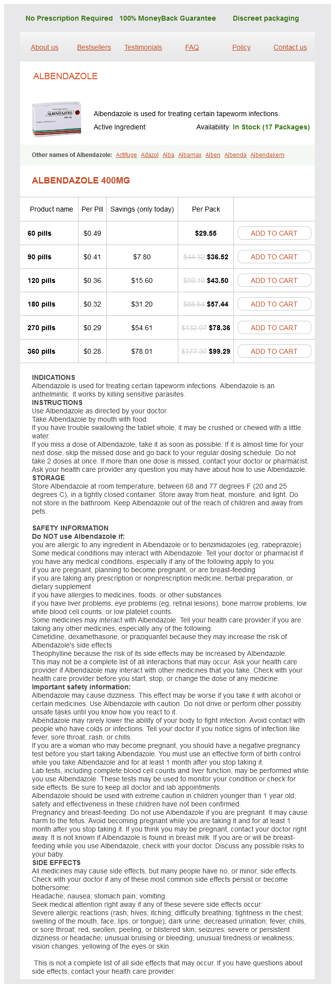
Albendazole Dosage and Price
Albendazole 400mg
- 60 pills - $29.55
- 90 pills - $36.52
- 120 pills - $43.50
- 180 pills - $57.44
- 270 pills - $78.36
- 360 pills - $99.29
Initially hiv infection rates by activity albendazole 400 mg otc, lymphoid hyperplasia develops as tubercles form, and then necrosis and caseation dominate. Early on, nodes are firm, discrete, and nontender, but with progression they tend to become matted and adherent to the overlying skin, which often becomes discolored, thickened, and scaly. Without treatment, spontaneous drainage ultimately occurs, leaving a draining sinus. Submandibular, submental, preauricular, anterior cervical, inguinal, or epitrochlear node may be the site of involvement, although cervical is most common. Increased pain and erythema, thinning of the overlying skin, and fluctuance on palpation signal that central necrosis has occurred. A, Early in the course of adenitis caused by Mycobacterium tuberculosis or atypical mycobacteria, enlargement of the node is gradual, tenderness is mild, and there is little or no sign of warmth or overlying inflammation. B, After several weeks the overlying skin becomes thickened, tense, discolored, and adherent to the node. Spontaneous resolution occurs over 12 to 24 months even in the absence of excision, making observation a reasonable course if removal is problematic due to nearby neurovascular structures. Cat-Scratch Disease Adenitis is a primary feature of cat-scratch disease, although lowgrade fever occurs in about 25% of patients. Ninety percent of patients have a history of either a cat scratch or contact with cats, especially kittens. Inoculation via cat scratch is the most common means of infection, but puncture wounds, flea bites, and dog scratches have also been implicated. Incidence is highest in fall and winter in temperate climates, with cases occurring with equal frequency year-round in tropical areas. Most patients are in the 5- to 14-year-old age range, but family clusters that include younger children and adults have been reported. Shortly thereafter, one or more regional nodes enlarge, becoming mildly painful and tender. Involved nodes are firm, and overlying warmth and mild redness may develop within a few days of enlargement. In order of frequency, axillary, cervical, submandibular, preauricular, epitrochlear, and inguinal nodes have been reported. With preauricular adenitis, associated conjunctivitis is common and suggests conjunctival inoculation as the source (a Parinaud syndrome). Discomfort generally subsides in 4 to 6 weeks, but the node may remain enlarged or may fluctuate in size for months. Diagnosis is made primarily by history, clinical picture, and course but can be confirmed by biopsy. Histology reveals granulomas with microabscesses, and Warthin-Starry silver stain may identify gram-negative pleomorphic coccobacilli. Serology is often positive, Adenitis Associated With Animal or Vector Contact In many children, acute local adenitis results from inoculation of a pathogen by an animal scratch or bite, from the bite of an insect vector transmitting a pathogen from an animal host, or from contact with a contaminated animal carcass. In some of these disorders, systemic symptoms are prominent; in others, the local adenitis is the primary manifestation. After the manifestations of local infection appear at the primary site (usually within 24 hours), a regional node becomes enlarged and tender. Although this illness is clinically indistinguishable from adenitis due to streptococci or staphylococci, P. An ulcerated papule, evident on his left cheek, was the site of a scratch inflicted by one of his kittens 2 weeks earlier. B, A line of papules is seen on the forearm of a 3-year-old at the site of a scratch inflicted by his new kitten 3 weeks before presentation. If lymph nodes are painful, suppurative aspiration can be performed and is preferable to incision and drainage because of concerns that the latter procedure may lead to prolonged drainage and scarring. The benefit of antimicrobial treatment in an immunocompetent host remains controversial. Tularemia Francisella tularensis most often produces an ulceroglandular syndrome in children, occurring in concert with systemic symptoms. Children may acquire disease from the bite of an arthropod vector, most commonly a tick; by handling or skinning dead animals; or after an animal bite (especially that of a cat that hunts rabbits). Onset is abrupt and characterized by fever, chills, headache, myalgias, vomiting, and possibly photophobia. A painful papule appears at the site of inoculation with subsequent axillary, epitrochlear, or inguinal adenitis. The involved regional node is firm and tender and may be associated with overlying erythema. Generalized adenopathy and hepatosplenomegaly may be noted in some patients; and in the second week of illness, a blotchy, erythematous maculopapular rash (or occasionally a vesicular, pustular, or nodosa exanthem) may appear. Without treatment, fever may persist for 2 to 3 weeks, and the ulcer may take as long as 1 month to heal. The diagnosis is suggested by history, geography, clinical picture, and course and may be confirmed serologically. If cultures are obtained, the microbiology laboratory should be notified as to the concern for tularemia. Rapid progression of systemic symptoms occurs, with the patient appearing toxic and apprehensive and often delirious with signs of neurologic dysfunction. If infection is suspected, the node should be aspirated to obtain material for culture, blood cultures should be performed, and broad-spectrum parenteral antibiotic therapy instituted. Parotitis Salivary glands can be inflamed due to viruses or, less frequently, bacteria. The most common viral etiologies associated with parotitis are mumps virus and enteroviruses, whereas the most common bacterial pathogen is S. Mumps (Epidemic Parotitis) Mumps is an acute viral illness that preferentially involves glandular and neural tissues.

ADDII BIOTECH is proud to announce that the company's manufacturing facility located at Baddii, Himachal Pradesh has recently been approved by Food & Drug Authority of Ghana.
Our Manufacturing capability includes a wide range of therapeutic products covering almost every segment.
ADDII BIOTECH given our strong emphasis on product quality and services.