
Atorvastatin
General Information about Atorvastatin
In conclusion, Atorvastatin has proven to be an efficient and widely-used medication for managing excessive cholesterol levels. It has numerous well being benefits and might play a significant position in decreasing the chance of heart illness and stroke. However, as with every medication, it is very important use it as prescribed and to seek the guidance of a healthcare professional earlier than making any changes to one's remedy plan. With correct use and life-style adjustments, Atorvastatin may help people maintain a wholesome heart and stay a longer, more healthy life.
Aside from its cholesterol-lowering effects, Atorvastatin additionally has a quantity of other well being advantages. Studies have discovered that it can reduce the chance of coronary heart attack, stroke, and even death in individuals with a history of cardiovascular disease. It has also been proven to improve cardiovascular health by lowering inflammation in the body and selling higher blood move.
There are certain factors that may improve an individual's risk of high ldl cholesterol, such as genetics, age, and food regimen. However, having excessive ldl cholesterol doesn't necessarily imply that a person is unhealthy. In reality, some folks may have excessive cholesterol due to inherited genetic factors that are beyond their control. For these people, Atorvastatin could be an efficient treatment to decrease their cholesterol levels.
One of the main concerns surrounding Atorvastatin is the price. Being a brand-name drug, it could be costlier than generic versions of statins. However, due to the expiration of its patent in 2012, generic variations of Atorvastatin are now available, making it extra reasonably priced for sufferers.
Atorvastatin is on the market in several strengths and formulations, ranging from 10 mg to 80 mg, and could be taken as a pill as quickly as a day. The dosage is set by the affected person's ldl cholesterol level and their general well being. It is important to note that Atorvastatin just isn't an various to a healthy life-style, together with a balanced diet and common train. It is supposed for use in conjunction with these way of life changes to effectively handle levels of cholesterol.
High cholesterol levels are a serious risk issue for heart disease and stroke, two of the leading causes of dying globally. Cholesterol is a waxy substance that may build up within the arteries, leading to blockages that can outcome in heart assaults and strokes. Atorvastatin works by reducing the quantity of cholesterol produced by the liver, thus reducing the buildup of plaque in the arteries and decreasing the risk of heart disease.
Atorvastatin, additionally known by its model name Lipitor, is a commonly prescribed medication used to decrease cholesterol and triglyceride levels in the physique. It belongs to a category of medicine called statins, which work by blocking an enzyme in the liver that's liable for producing ldl cholesterol. Atorvastatin is one of the most widely prescribed medications for top ldl cholesterol, with an estimated 21 million prescriptions written in the United States alone in 2019.
Atorvastatin is mostly well-tolerated, with mild unwanted side effects such as headache, muscle ache, and diarrhea being reported by some users. In uncommon circumstances, more critical unwanted side effects similar to liver harm and muscle breakdown could occur. It is essential to consult a healthcare skilled before beginning Atorvastatin or another medicine, as they'll assess the individual's medical history and decide the appropriate dosage and monitoring plan.
S haematobium is associated with lesions of the lower genital tract (vulva, vagina, and cervix) in women, prostatitis and hematospermia in men, and certain forms of bladder cancer cholesterol medication mechanism of action generic atorvastatin 40 mg buy. Other organ systems can be involved; for example, eggs can embolize in the lungs, causing pulmonary hypertension. Less commonly, eggs can lodge in the central nervous system, causing severe neurologic complications. These larvae can penetrate human skin but eventually die in the dermis and do not cause systemic disease. Skin manifestations include pruritus at the penetration site a few hours after water exposure, followed in 5 to 14 days by an intermittent pruritic, sometimes papular, eruption. Etiology the trematodes (flukes) S mansoni, S japonicum, Schistosoma mekongi, and Schistosoma inter calatum cause intestinal schistosomiasis, and S haematobium causes urogenital disease. After development and asexual replication in snails, cercariae emerge and penetrate the skin of humans in contact with water. Children are commonly first infected when they accompany their mothers to lakes, ponds, and other open freshwater sources. School-aged children are typically the most heavily infected people in the community because of prolonged wading and swimming in infected waters. They are also important in maintaining transmission through behaviors such as uncontrolled defecation and urination. Communicability lasts as long as infected snails are in the environment or live eggs are excreted in the urine and feces of humans into freshwater sources with appropriate snails. S mansoni occurs throughout tropical Africa, in parts of several Caribbean islands, and in areas of Venezuela, Brazil, Suriname, and the Arabian Peninsula. Thus, schistosomiasis can be diagnosed in patients many years after they have left an area with endemic infection. Incubation Period Approximately 4 to 6 weeks for S japonicum, 6 to 8 weeks for S mansoni, and 10 to 12 weeks for S haematobium. Diagnostic Tests Eosinophilia is common and may be intense in Katayama fever (acute schistosomiasis). Infection with S mansoni and other species (except S haematobium) is determined by microscopic examination of stool specimens to detect characteristic eggs, but results may be negative if performed too early in the course of infection. In light infections, several stool specimens examined by a concentration technique may be needed before eggs are found, or eggs may be seen in a biopsy of the rectal mucosa. Serologic tests, available through the Centers for Disease Control and Prevention and some commercial laboratories, can be helpful for detecting light infections. However, results of these antibody-based tests remain positive for many years and are not useful in differentiating ongoing infection from past infection or reinfection. A skin biopsy may demonstrate larvae, but their absence does not exclude the diagnosis. Treatment the drug of choice for schistosomiasis caused by any species is praziquantel; the alternative drug for S mansoni is oxamniquine, although this drug is not available in the United States (it is used in some areas of Brazil). Praziquantel does not kill developing worms; therapy given within 4 to 8 weeks of exposure should be repeated 1 to 2 months later to improve the rate of parasitologic cure. Once inside the host, the S haematobium parasite passes through 2 developmental generations of sporocysts and is released by the snail into its environment as cercariae. The stages in the snail include 2 generations of sporocysts (4) and the production of cercariae (5). On release from the snail, the infective cercariae swim, penetrate the skin of the human host (6), and shed their forked tail, becoming schistosomulae (7). The schistosomulae migrate through several tissues and stages to their residence in the veins (10). Adult worms in humans reside in the mesenteric venules in various locations, which, at times, seem to be specific for each species (10). For instance, Schistosoma japonicum is more frequently found in the superior mesenteric veins draining the small intestine (A), and Schistosoma mansoni occurs more often in the superior mesenteric veins draining the large intestine (B). However, both species can occupy either location, and they are capable of moving between sites, so it is not possible to state unequivocally that one species only occurs in one location. Schistosoma haematobium most often occurs in the venous plexus of the bladder (C), but it can also be found in the rectal venules. The females (size 720 mm; males slightly smaller) deposit eggs in the small venules of the portal and perivesical systems. The eggs are moved progressively toward the lumen of the intestine (S mansoni and S japonicum) and of the bladder and ureters (S haematobium) and are eliminated with feces or urine, respectively (1). Pathology of S mansoni and S japonicum schistosomiasis includes Katayama fever, presinusoidal egg granulomas, Symmers pipestem periportal fibrosis, portal hypertension, and occasional embolic egg granulomas in the brain or spinal cord. Pathology of S haematobium schistosomiasis includes hematuria, scarring, calcification, squamous cell carcinoma, and occasional embolic egg granulomas in the brain or spinal cord. On release from the snail host, the infective cercariae swim, penetrate the skin of the human host, and shed their forked tail, becoming schistosomulae. The schistosomulae migrate through several tissues and stages to their residence in the veins. This micrograph reveals signs of schistosomiasis infection of the liver, also known as pipestem cirrhosis (magnification x500). Pipestem cirrhosis occurs when schistosomes infect the liver (ie, hepatic schistosomiasis), which causes scarring to occur, thereby entrapping parasites and their ova in and around the hepatic portal circulatory vessels.
Hypothalamic neuropeptide Y inhibits gastric acid output in rat: role of the autonomic nervous system cholesterol ranges for male buy atorvastatin no prescription. Neurochemistry of superficial spinal neurones projecting to nucleus of the solitary tract that express c-fos on chemical somatic and visceral nociceptive input in the rat. Immunohistochemical profiles of spinal lamina I neurones retrogradely labelled from the nucleus tractus solitarii in rat suggest excitatory projections. Gastric dysmotility after abdominal surgery in persons with cervical spinal cord injury: a case series. Response properties of antral mechanosensitive afferent fibers and effects of ionotropic glutamate receptor antagonists. Vagal and spinal mechanosensors in the rat stomach and colon have multiple receptive fields. Electrophysiological characterization of vagal afferents relevant to mucosal nociception in the rat upper oesophagus. Sensitivity of vagal mucosal afferents to cholecystokinin and its role in afferent signal transduction in the rat. Altered mechanosensitive properties of vagal afferent fibers innervating the stomach following gastric surgery in rats. Esophageal distension induced gastric relaxation is mediated in part by vagal peripheral reflex mechanism in rats. Noradrenergic neurons in the rat solitary nucleus participate in the esophageal-gastric relaxation reflex. Neurotransmitters of the non-adrenergic non-cholinergic relaxation of proximal stomach. Relationships between the morphology and function of gastric and intestinal distention-sensitive neurons in the dorsal motor nucleus of the vagus. Impact of antral mechanoreceptor activation on the vago-vagal reflex in the rat: functional zonation of responses. Vagal afferent discharge from mechanoreceptors in different regions of the ferret stomach. Effect of duodenal distension on the pyloric sphincter and antrum and the gastric corpus: duodenopyloric reflex. The vagal control of the pyloric motor function: a physiological and immunohistochemical study in cat and man. Gastric distension-induced pyloric relaxation: central nervous system regulation and effects of acute hyperglycaemia in the rat. Vagal afferent stimulation-evoked gastric secretion suppressed by paraventricular nucleus lesion. Vagal influences on gastric acid secretion in response to gastric distension in the ferret. Capsaicin-sensitive vagal afferent fibers and stimulation of gastric acid secretion in anesthetized rats. Secretin activates vagal primary afferent neurons in the rat: evidence from electrophysiological and immunohistochemical studies. Peripheral secretin-induced Fos expression in the rat brain is largely vagal dependent. Duodenal lipid inhibits gastric acid secretion by vagal, capsaicin-sensitive afferent pathways in rats. Characterization of saturable binding sites for circulating pancreatic polypeptide in rat brain. Vagal control of digestion: modulation by central neural and peripheral endocrine factors. Proteinaseactivated receptors in the nucleus of the solitary tract: evidence for glial-neural interactions in autonomic control of the stomach. Morphology of identified preganglionic neurons in the dorsal motor nucleus of the vagus. Norepinephrine effects on identified neurons of the rat dorsal motor nucleus of the vagus. Morphological differences between planes of section do not influence the electrophysiological properties of identified rat dorsal motor nucleus of the vagus neurons. Anatomical demonstration of vagal input to nicotinamide acetamide dinucleotide phosphate diaphorase-positive (nitrergic) neurons in rat fundic stomach. Cholecystokinin activates catecholaminergic neurons in the caudal medulla that innervate the paraventricular nucleus of the hypothalamus in rats. Electrophysiological and morphological heterogeneity of rat dorsal vagal neurones which project to specific areas of the gastrointestinal tract. Adenosine-induced inhibition of vagal motoneuron excitability: receptor subtype and mechanisms. Response of neurons in the dorsal motor nucleus of the vagus to thyrotropin-releasing hormone. Dorsal motor nucleus of the vagus nerve: a cyto- and chemoarchitectonic study in the human. Dorsal medullary serotonin and gastric motility: enhancement of effects by thyrotropin-releasing hormone. Discharge of efferent vagal fibers supplying gastric antrum: indirect study by nerve suture technique. Representation of the cecum in the lateral dorsal motor nucleus of the vagus nerve and commissural subnucleus of the nucleus tractus solitarii in rat. The dorsal vagal complex forms a sensory-motor lattice: the circuitry of gastrointestinal reflexes.
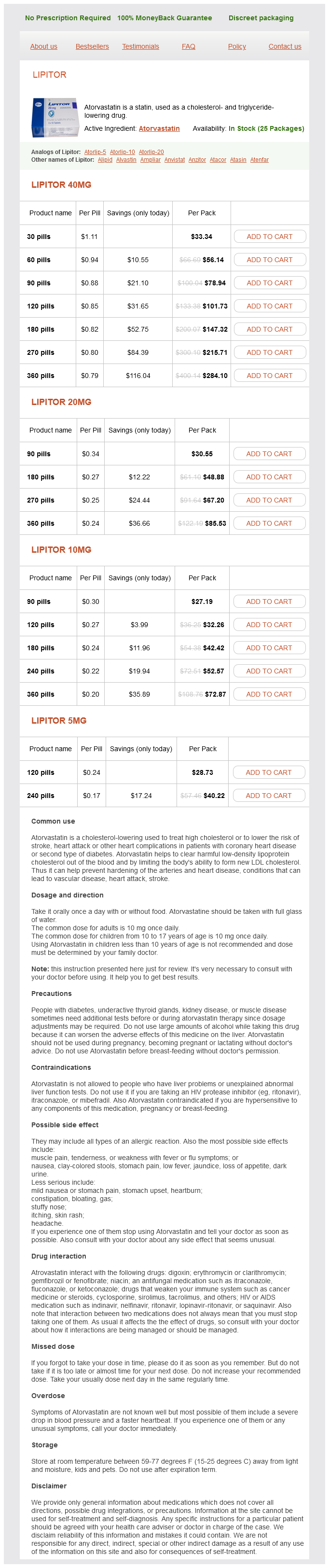
Atorvastatin Dosage and Price
Lipitor 40mg
- 30 pills - $33.34
- 60 pills - $56.14
- 90 pills - $78.94
- 120 pills - $101.73
- 180 pills - $147.32
- 270 pills - $215.71
- 360 pills - $284.10
Lipitor 20mg
- 90 pills - $30.55
- 180 pills - $48.88
- 270 pills - $67.20
- 360 pills - $85.53
Lipitor 10mg
- 90 pills - $27.19
- 120 pills - $32.26
- 180 pills - $42.42
- 240 pills - $52.57
- 360 pills - $72.87
Lipitor 5mg
- 120 pills - $28.73
- 240 pills - $40.22
In rodents, this information ascends to higher centers through second-order neurons in the pons (PbN, solid line) total cholesterol medical definition cheap atorvastatin 5 mg visa. Gustatory axons of the vagus nerve enter the medulla immediately caudal to the facial and glossopharyngeal fibers with less overlap. In rodents, the most intense taste responses occur posteriorly in the medial parabrachial nucleus, which is a densely packed layer of cells just ventral to the brachium conjunctivum. The parabrachial nuclei are unique among brainstem sensory relays in that they project to both the dorsal thalamus and the limbic system. In monkeys, the projections of the parabrachium resemble those of the rat, but are most likely a general visceral rather than a gustatory relay. It appears that in primates the parabrachium is not an obligatory relay for ascending gustatory information as it is in rodents. Interspecies differences within the parabrachium have little bearing on the locus of the gustatory area of the thalamus. The thalamic gustatory relay occupies homologous structures in all mammals examined, including rat, rabbit, dog, hamster, and monkey. This subnucleus also contains, in addition to gustatory neurons, neurons that respond to tactile and/or thermal stimuli. In humans, axons from these thalamic neurons travel through the ipsilateral posterior limb of the internal capsule to terminate in the inner portion of the frontal operculum and anterior insular cortex and in the rostral extension of Brodmann area 3B on the lateral convexity of the postcentral gyrus. In primates, an additional region of cortex, the lateral posterior orbitofrontal cortex, receives input from the primary taste cortex and acts as a site of integration for taste, olfactory, somatosensory, and visual cues thought important in appreciation of flavor, food reward, and control of feeding. Some speculate that this anatomical locus may be the neural substrate for flavor perception. There are also reflex connections within the brainstem that involve oromotor nuclei, adjacent premotor reticular formation centers, and salivatory nuclei. More rostral gustatory nuclei tend to have more connections with reticular formation and anterior oromotor centers; more caudal gustatory nuclei have more substantial connections with oropharyngeal motorneurons of the nucleus ambiguous. Another distinctive feature of this cell type is the presence of apically located electrondense granules (100400 nm). These granules are thought to exocytose into the taste pore, although their content or function remains unknown. It remains uncertain whether type I cells express taste receptor molecules that detect taste stimuli. They are spherical in shape and are found at the basal end of the taste bud, close to the basement membrane. The mature taste bud expresses many of the same signaling markers observed in development, suggesting some similarity of the processes of adult regeneration to embryonic development. For example, as is observed in the developing tongue, the protein sonic hedgehog (Shh) is also expressed in the adult taste bud in basal cells and its receptor patched (Ptc-1) in the epithelial region adjacent to the bud. Expression of the Shh target gene Gli1 was observed in the same region where Ptc-1 is expressed, indicating these cells respond to Shh. After denervation of the glossopharyngeal nerve, Shh expression decreased immediately, perhaps due to the neural loss, with subsequent Ptc-1 loss following. Sox2 is also highly expressed in basal epithelial cells of the adult tongue, as well as in development, and its expression in the adult is also dependent upon neural innervation. Sox2 is thought to be required for development of the taste bud and may play an important role in its continued maintenance. Their cytoplasm is moderately translucent and this cell type has sometimes been referred to as an intermediate cell to distinguish it from light and dark cells. These cells do, however, express transduction components that appear essential for sour taste transduction. Traditionally, taste buds had been viewed as consisting of two functional cell types: receptor cells, which had synapses with afferent nerve fibers, and supporting cells. These studies produced initial confusion when the presence of multiple signaling agents, both classic neurotransmitters and neuropeptides, became evident within the bud. Collectively, these findings led to the classification of transduction mechanisms within the bud into early and late events. Late transduction mechanisms, on the other hand, describe the processing of information among cells of the taste bud by cell-to-cell communication, which ultimately shapes the neural discharge. These events include lateral inhibition, gain modulation, and, possibly, adaptation. Thus single cells within the bud are influenced not only through apical receptor activation, but also through basolateral neurotransmitter release. At present, late transduction events are not well understood and how these hardwired pathways participate in the transduction cascades of taste quality processing is the subject of active investigation. T1Rs have a characteristic long extracellular C-terminus, whereas T2R receptors lack this domain. Salty and sour stimuli are transduced by ionotropic receptors that allow direct entry of the sodium or hydrogen cation into the cell thus depolarizing it. Sweet, bitter, and umami stimuli are transduced by metabotropic receptors that fall into two families: T1R and T2R (or Tas1 and Tas2). The T1R family consists of three members (T1R1, T1R2, and T1R3) that function as two sets of dimers. The T1R1/T1R3 dimer responds to amino acids, while the T1R2/T1R3 dimer responds to sweet stimuli. The T2R family, on the other hand, consists of about 30 members that respond to bitter stimuli. Both families of taste receptors are transduced by the same intracellular cascade. Many speculated that the genes of these receptors would map onto the sac locus, a region on distal chromosome 4 previously identified by genetic studies to be involved with sweet taste in mice.

ADDII BIOTECH is proud to announce that the company's manufacturing facility located at Baddii, Himachal Pradesh has recently been approved by Food & Drug Authority of Ghana.
Our Manufacturing capability includes a wide range of therapeutic products covering almost every segment.
ADDII BIOTECH given our strong emphasis on product quality and services.