
Aygestin
General Information about Aygestin
It is essential to inform your physician of any other medicines you're taking earlier than beginning Aygestin. This includes over-the-counter drugs, vitamins, and herbal supplements. Some medications might work together with Aygestin and have an effect on its effectiveness.
Abnormal bleeding is usually a results of hormonal imbalances or underlying medical situations similar to fibroids or endometriosis. Aygestin works by mimicking the hormone progesterone within the physique, which helps to stability out the levels of estrogen and progesterone. This helps to regulate the menstrual cycle and reduce heavy bleeding.
In rare cases, Aygestin could enhance the chance of blood clots, especially in ladies who smoke, are over the age of 35, or have a history of blood clots. Therefore, you will want to focus on any potential threat components together with your doctor before starting Aygestin.
As with any medication, there are potential unwanted effects related to Aygestin. These might include nausea, headache, breast tenderness, and changes in menstrual bleeding patterns. If any of these unwanted effects persist or become bothersome, it could be very important seek the assistance of together with your physician.
In addition, Aygestin shouldn't be used by pregnant girls as it might harm the unborn child. It is also not beneficial to be used while breastfeeding, as small amounts of the medicine might move into breast milk.
Aside from its use in managing menstrual and uterine problems, Aygestin can be used for other circumstances as determined by a doctor. These may embody treating irregular bleeding brought on by hormonal imbalances, preventing being pregnant, or managing signs of premenstrual syndrome (PMS).
In conclusion, Aygestin is a drugs that's used to deal with sure menstrual and uterine problems. It works by regulating the menstrual cycle, managing heavy bleeding, and reducing symptoms of endometriosis. It is important to observe the prescribed dosage and to discuss any potential risks together with your physician before starting this medicine. With correct use, Aygestin can present aid and improve the standard of life for girls with these situations.
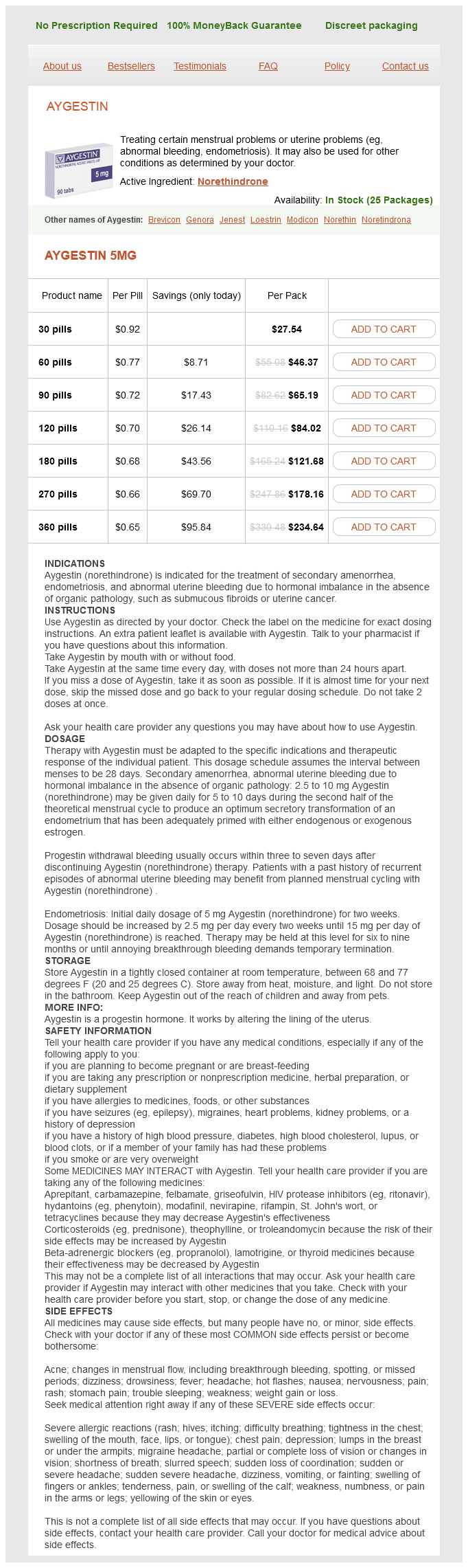
Aygestin, also known as norethindrone, is a medication used for the treatment of certain menstrual and uterine problems. These circumstances may cause disruptions in a woman’s every day life and overall well being, and Aygestin offers relief by regulating the menstrual cycle and managing irregular bleeding.
In women with endometriosis, Aygestin works by suppressing the expansion of the endometrial tissue, which can cause painful periods and infertility. It additionally reduces the inflammation and swelling related to endometriosis, offering relief from signs corresponding to pelvic ache and discomfort.
Aygestin is on the market in pill kind and is usually taken as soon as a day, with or without meals. It is essential to take the medication at the same time every day to keep up constant ranges within the body. It can be important to comply with the dosage prescribed by your physician and not to miss any doses.
Injury of the spinal cord due to rupture of an intervertebral disc during muscular efort women's health bendigo hospital discount generic aygestin uk. Spinal compression caused by enchondrosis of the intervertebral ibrocartilage with a review of the recent literature. Complications in thoracoscopic spinal surgery: a study of 90 consecutive patients. A calciied thoracic intervertebral disk with herniation and spinal cord compression in a child: case report. Multiple protrusions of intervertebral disks in the upper thoracic region: report of case. Chapter 45 Thoracic Disc Disease lumbar spine: insights into the etiopathogenesis of spinal degeneration. Protrusion of intervertebral discs: study of their distribution, characteristics and efects on the nervous system. Asymptomatic versus symptomatic herniated thoracic discs: their frequency and characteristics as detected by computed tomography ater myelography. Post-operative pain-related morbidity: video-assisted thoracic surgery versus thoracotomy. Angiographic study of the efect of laminectomy in the presence of acute anterior epidural masses. Der horakale Bandscheibenprolaps als extramedullarer Spinaltumor und in seinen Beziehungen zu internen Organsyndromen. Intradural disc herniations in the cervical, thoracic, and lumbar spine: report of three cases and review of the literature. Deleterious efects of discography radiocontrast solution on human annulus cell in vitro: changes in cell viability, proliferation, and apoptosis in exposed cells. Results of surgical treatment for thoracic myelopathy: minimum 2-year follow-up study in 132 patients. Chylorrhea: a rare complication of thoracoscopic discectomy of the thoracolumbar junction. Lateral extracavitary approach to the spine for thoracic disc herniations: report of 23 cases. Surgical treatment for central calciied thoracic disk herniation: a novel L-shaped osteotomy. Clinical analysis of video-assisted thoracoscopic spinal surgery in the thoracic or thoracolumbar spinal pathologies. Analysis of hard thoracic herniated discs: review of 18 cases operated by thoracoscopy. Video-assisted thoracoscopic surgery under O-arm navigation system guidance for the treatment of thoracic disk herniations: surgical techniques and early clinical results. Mini-thoracotomy or thoracoscopic treatment for medially located thoracic herniated disc Techniques for the operative management of thoracic disc herniation: minimally invasive thoracic microdiscectomy. Pain can originate from several anatomic structures within the spine, making it diicult for both the patient and physician to localize. Degenerative disc disease also contributes to the pathogenesis of secondary spinal disorders such as spinal stenosis and degenerative spondylolisthesis. Only 20% of patients still disabled ater 1 year will return to work, and only 2% return ater 2 years. During the instability phase abnormal motion can occasionally be seen on lexion-extension radiographs; however, the spinal segments during this phase of degeneration more oten show no demonstrable radiographic instability. Although a deining characteristic of the cells is the presence of numerous cytoplasmic processes, a remarkable inding is that the cells contain few, if any, mitochondria. Moreover, since the nucleus pulposus has no blood supply, the oxygen tension within the disc is very low. Because of the presence of hydrophilic aggrecan, the disc exhibits a high osmotic pressure. Although the movement capable of a single spinal motion segment is quite limited, the range of motion supported by the 24 presacral motion segments of the spine in its entirety is remarkable. Found between two rigid vertebral bodies, the disc allows this movement while simultaneously supporting compressive loads. Major characteristics of these tissue components of the disc are highlighted below. Nucleus Pulposus he central nucleus pulposus is derived from the embryonic notochord, and notochordal cells remain in the tissue ater birth and into adult life. During development, the nucleus is highly cellular; ater birth and an initial period of cell growth, the number of cells declines. Among the large notochordal cells, much smaller cells, possibly derived from the notochordal sheath, can also be seen. Neither necrotic nor apoptotic Compressive deformation of the nucleus pulposus is limited circumferentially by the anulus ibrosus, which experiences the compressive loads of the torso as hoop stress. Since the anulus ibrosus experiences tensile stress as opposed to compression, its anatomy and biochemical composition are distinct from the nucleus pulposus. Many consider the inner anulus a transition zone because it difers substantially from the outer region. Compared with the outer anulus ibrosus, where the cells are elongated and fusiform aligned with the long axis of the ibrils, the cells of the inner anulus are spherical in shape and more closely resemble chondrocytes.
It serves as an interface between the sot nucleus pulposus and the dense bone of the vertebrae as well as a biomechanical barrier that prevents the disc from applying pressure directly to the bone women's health center towson md order 5 mg aygestin with amex. In addition, the end plate cartilage plays a role in maintaining nucleus pulposus cell viability and controlling biosynthetic activities. Although the cells do not undergo terminal diferentiation, collagen X may be present in the central region of the end plate, perhaps in relationship to focal areas of endochondral bone formation. Work in rabbit and rat models suggests that end plate chondrocytes may contribute a cell fraction that makes up some of the nucleus pulposus and the anulus ibrosus in normal development and during injury. In his review on the end plate, Moore28 noted that vascular channels penetrate the cartilage during development, but at maturity the vessels become narrow or even obliterated. It is likely that this change impacts the nutrient supply to both the cartilage and the disc. Supporting this, Crock and Yoshizawa30 showed that the central region of the end plate, where there is a high concentration of channels, is freely permeable to small molecules. Clinically, it is not uncommon to note that the central region undergoes sclerosis or mineralization with alterations in the mechanical properties of the cartilage. When this occurs, nucleus pulposus tissue can be forced through the end plate into the underlying bone of the vertebrae. Blood vessels originating in the vertebral body traverse the supericial region of the end plates, but none of these vessels iniltrates the nucleus pulposus. Gruber and colleagues31 showed that the anulus is avascular except for small capillary beds in the dorsal and ventral surfaces, and this vasculature never enters the nucleus pulposus. Experimental measurements done in canine discs34 as well as modeling studies by Bartels et al. Critical functions include energy metabolism, angiogenesis, cell survival, autophagy and apoptosis, matrix synthesis, proliferation, self-renewal and diferentiation, radical dismutation, and pH regulation. Many of these functions are critical for survival and functioning of the nucleus pulposus cells in the avascular niche of the intervertebral disk. Eventually the nucleus pulposus is replaced with a ibrocartilaginous tissue with inferior biomechanical properties. Ishihara and colleagues were the irst to demonstrate this relationship in disc tissue using bovine nucleus pulposus explants. For detailed discussion of osmosensing in intervertebral disc see recent review by Johnson et al. It is composed of a sensory branch from the ventral root and a sympathetic branch from the gray rami communicans near the distal pole of the dorsal root ganglion. Alterations in gene expression and transcription factors may be responsible for cell senescence within the disc. Both anabolic and catabolic processes are upregulated during the early stages of degeneration; however, anabolic repair processes fail to keep up with the catabolic processes and matrix degeneration ensues over time. As the process progresses, the inner layers of the anulus and the nucleus pulposus gradually become indistinguishable and change into a stif desiccated ibrocartilaginous material. Loss of end plate vascularity and porosity leads to a reduction in the inlux of nutrients and elux of waste products. Lactate levels rise locally within the hypovascular disc secondary to increased production and decreased removal. Cell apoptosis occurs as a result of decreased tissue pH,66 and the biosynthetic reparative capability of the disc is thus further impaired. Focal elevations in shear stresses at the disc level may further adversely afect the disc structure and result in anular damage. Eventually communicating channels may develop between the peripheral layers of the anulus and the nucleus, and disc material can then herniate through these issures. Changes in disc structure alter the loading response and alignment of the spinal column. However, pain does not always correlate with morphologic changes in the disc and mechanical compression. Alteration in loading patterns between the end plate and disc leads to anular damage and the potential for herniation. Perturbation of the disc also leads to degeneration and pain in other segmental structures such as the facet joints, ligaments, and paraspinal muscles. Studies have implicated occupations that require repetitive liting or pulling, prolonged sitting76 (such as motor vehicle driving77), and whole-body vibration. Prolonged mechanical load can therefore cause a disruption of difusion that may accelerate disc degeneration; however, this hypothesis is yet to be conirmed clinically. In the upper lumbar spine only 7% of the variability was explained by occupation, 16% by age, and 77% by familial aggregation. In the lower lumbar spine recreational physical loading explained 2% of variability, age explained 9%, and familial aggregation explained 43%. It is important to note that there is a growing consensus spearheaded by Carragee that discography may accelerate disc degeneration, loss of disc height, and overall progression of pathology and symptoms. Two potential sources that have been implicated as contributors to discogenic pain are sensitization of nerve endings by release of chemical mediators and neurovascular ingrowth into the degenerated disc. Radial anular tears provide a route for nuclear material and noxious chemicals to leak from the disc and contact the dural sac and nerve roots; some studies have shown that autologous nucleus pulposus alone has the capacity to produce a robust inlammatory response. Subclinical Bacterial Infection of the Spinal Motion Segment as a Possible Initiator of Low Back Pain here is growing interest and controversy in the notion that subclinical anaerobic bacterial infection could play a role in symptomatic disc degeneration. Type 1 changes were described as decreased signal intensity on T1-weighted images with increased signal intensity on T2-weighted images; type 2 changes were described as increased signal intensity on T1-weighted images with slightly increased intensity on T2-weighted images. Fat appears bright and increases the signal intensity on T1-weighted images, whereas water increases signal intensity on T2-weighted images. Consequently, painful type 1 changes are associated with edema in the vertebral bodies. Simple environmental contamination would not likely favor anaerobic bacteria over the more commonly aerobic skin lora.
Aygestin Dosage and Price
Aygestin 5mg
- 30 pills - $27.54
- 60 pills - $46.37
- 90 pills - $65.19
- 120 pills - $84.02
- 180 pills - $121.68
- 270 pills - $178.16
- 360 pills - $234.64
The use of segmental pedicle screw ixation for the posterior treatment of pediatric and adult idiopathic scoliosis curves has become the primary instrumentation construct menstrual smell purchase aygestin pills in toronto. In addition, thorough bone grafting with a combination of autogenous bone, allograft bone, and/or the use of osteobiologics, especially in the adult population, has become routine at many centers throughout North America. Optimal surgical outcomes in the treatment of idiopathic scoliosis deformities include proper patient selection, exacting surgical technique, and a well-balanced spinal alignment with minimal to no complications. Summary Understanding and treatment of spinal deformities has broadened; however, idiopathic scoliosis remains a diagnosis of exclusion. With advances in genetic mapping of idiopathic scoliosis, better understanding of the etiology and incidence of the disease is promising. It is hoped that better understanding will bring earlier identiication, more insight into curve progression risk, and treatments of the condition without the need for major surgery of severe curves. Possible treatment modalities include close observation, bracing, and surgical intervention. Selective fusions should be performed whenever possible, and critical curve analysis should be performed preoperatively with all available objective modalities. Direct vertebral rotation ofers improved thoracic correction and a decreased need for thoracoplasty. Complex, decompensated, large, rigid curves and curves previously fused may require osteotomies to achieve the desired correction. Some pitfalls of scoliosis surgery, such as decompensation and adding on of a fused curve, can be avoided when these principles are applied. One must be mindful of the lessons of the past in understanding the assessment and management of spinal deformity. Spinal surgeons constantly must strive for improvements in surgical technique that lead to shorter, selective fusions and a balanced spine with maximum possible correction. Safety for patients is of the utmost importance and is achieved by appropriate training, careful patient selection, and adherence to the principles of deformity surgery. Patient evaluation skills and highly specialized technical skills are essential for the scoliosis surgeon. Adjuncts to posterior correction possibly can help obviate more extensive approaches. Avoidance and treatment of complications in the preoperative, intraoperative, and postoperative periods is important. Some form of spinal cord monitoring is mandatory for all scoliosis corrective procedures. Intraobserver and interobserver reliability of the classiication of thoracic adolescent idiopathic scoliosis. This study showed poor to fair reliability of the King classiication of adolescent idiopathic scoliosis, questioning its usefulness as an accurate system. Curve prevalence of a new classiication of operative adolescent idiopathic scoliosis: does classiication correlate with treatment Selective anterior fusion of thoracolumbar/lumbar curves in adolescents: When can the associated thoracic curve be left unfused Selective thoracic fusion for adolescent idiopathic scoliosis with C modiier lumbar curves: 2- to 16- year radiographic and clinical results. Satisfactory results were achieved with selective thoracic fusion of properly selected C modiier lumbar curves with under-correction of the instrumented thoracic curve (36%) to match the spontaneous correction of the lumbar curve (34%). Idiopathic scoliosis: the prognosis, diagnosis, and operative indications related to curve patterns and the age at onset. Molecular determinants of melatonin signaling dysfunction in adolescent idiopathic scoliosis. A postural dysequilibrium as an etiological factor in idiopathic scoliosis [abstract O]. In Programs and Abstracts of the 17th Annual Meeting of the Scoliosis Research Society, Denver, 1982, p 52. Genetic linkage localizes an adolescent idiopathic scoliosis and pectus excavatum gene to 18q. Long-term observation and management of resolving infantile idiopathic scoliosis: a 25-year follow-up. In Programs and Abstracts of the 40th Annual Meeting of the Scoliosis Research Society, Miami, 2005, p 120. Selective anterior fusion of thoracolumbar/lumbar curves in adolescents: When can the associated thoracic curve be let unfused In the 8th Proceedings of the International Congress on Cotrel-Dubousset Instrumentation. Efectiveness of treatment with a brace in girls who have adolescent idiopathic scoliosis: a prospective, controlled study based on data from the Brace Study of the Scoliosis Research Society. Eicacy of exercise therapy for the treatment of adolescent idiopathic scoliosis: a review of the literature. Shoulder balance ater surgery in patients with Lenke type 2 scoliosis corrected with the segmental pedicle screw technique. Incidence of neural axis abnormalities in infantile and juvenile patients with spinal deformity: Is a magnetic resonance image screening necessary Relationship of syrinx size and tonsillar descent to spinal deformity in Chiari malformation type I with associated syringomyelia. Validation of radiographic sotware to determine Lenke classiication [abstract 54]. In Programs and Abstracts of the 40th Annual Meeting of the Scoliosis Research Society, Miami, 2005, p 93. Adolescent idiopathic scoliosis: a new classiication to determine extent of spinal arthrodesis.

ADDII BIOTECH is proud to announce that the company's manufacturing facility located at Baddii, Himachal Pradesh has recently been approved by Food & Drug Authority of Ghana.
Our Manufacturing capability includes a wide range of therapeutic products covering almost every segment.
ADDII BIOTECH given our strong emphasis on product quality and services.