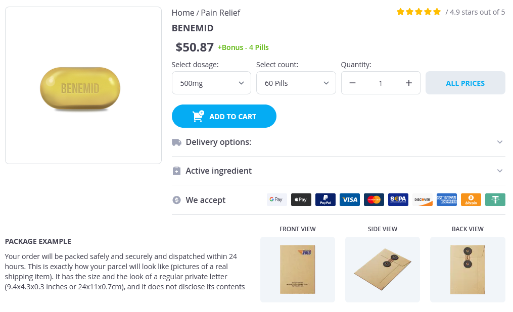
Benemid
General Information about Benemid
In addition to treating gout, Benemid has additionally been discovered to be effective in the remedy of other situations similar to sure forms of kidney stones and a rare genetic disorder called familial juvenile hyperuricemic nephropathy. It has also been used off-label for the remedy of certain infections and to extend the plasma levels of sure antibiotics.
Gout is a typical form of arthritis that involves sudden, extreme assaults of pain, redness, swelling, and tenderness within the joints. It is attributable to excessive ranges of uric acid within the body, which may crystallize and form deposits in the joints, leading to painful flare-ups. Though there are numerous treatments obtainable for gout, one medication particularly stands out for its capability to stop the formation of uric acid - Benemid.
Benemid is primarily used in patients who cannot take different medications corresponding to allopurinol or febuxostat, that are generally prescribed for gout, as a end result of both unwanted effects or allergic reactions. It can be utilized in combination with these medications for sufferers who do not respond properly to them. In addition, Benemid is used as a prophylactic treatment to forestall recurrent gout assaults.
In conclusion, Benemid is a useful treatment for the therapy of gout and different conditions attributable to excessive ranges of uric acid within the physique. It provides an alternative possibility for many who cannot take different drugs for gout and has been proven to be efficient in lowering uric acid ranges. However, as with every medicine, it is necessary to use Benemid as prescribed and under the steerage of a healthcare professional.
Benemid is on the market in both tablet and injectable form. The beneficial dose is normally 250 mg twice a day, although this will likely differ relying on the severity of the condition and response to therapy. It is important to note that whereas Benemid might help decrease uric acid levels, it does not present immediate aid from gout symptoms. Therefore, different medicines might have to be prescribed for acute relief throughout gout attacks.
Like any treatment, Benemid is not with out its unwanted facet effects. The commonest side effects embody abdomen upset, headache, dizziness, and skin rash. In rare instances, it may also cause more critical unwanted effects like kidney stones, low platelet rely, and blood problems. As with any medication, it is very important consult with a physician earlier than beginning Benemid and report any unwanted aspect effects experienced.
Benemid, also recognized as probenecid, is an anti-gout agent that has been used for over 60 years to deal with symptomatic hyperuricemia, a situation in which there is extra uric acid within the body. It works by blocking the reabsorption of uric acid in the kidneys, permitting the excess uric acid to be excreted through urine. This results in a decrease within the general levels of uric acid within the body and reduces the chance of gout attacks.
Determination of the degree of morphological regression following chemotherapy in malignant bone tumors pain treatment hemorrhoids 500 mg benemid for sale. Osteosarcoma: relationship of response to preoperative chemotherapy and type of surgery to local recurrence. Long-term follow-up of patients with doxorubicin-induced cardiac toxicity after chemotherapy for osteosarcoma. The effect of intra-arterial versus intravenous cisplatinum in the neoadjuvant treatment of osteosarcoma of the limbs: the experience at the Rizzoli Institute. Influence of methotrexate dose intensity on outcome of patients with high grade osteogenic osteosarcoma. Influence of methotrexate dose intensity on outcome of patients with high grade osteogenic osteosarcoma: analysis of the literature. Chemotherapy for osteosarcoma without high-dose methotrexate: a 12-year follow-up on 53 patients. Long-term outcome for patients with nonmetastatic osteosarcoma of the extremity treated at the Istituto Ortopedico Rizzoli according to the Istituto Ortopedico Rizzoli/Osteosarcoma-2 Protocol: an updated report. Received dose and dose-intensity of chemotherapy and outcome in nonmetastatic extremity osteosarcoma. High-dose chemotherapy in the treatment of relapsed osteosarcoma: an Italian sarcoma group study. Interferon-alpha as the only adjuvant treatment in high-grade osteosarcoma: long term results of the Karolinska Hospital series. Neoadjuvant chemotherapy for osteosarcoma of the extremities with metastases at presentation: recent experience at the Rizzoli Institute in 57 patients treated with cisplatin, doxorubicin, and a high dose of methotrexate and ifosfamide. Metastatic osteosarcoma at diagnosis: prognostic factors and longterm outcome-the French pediatric experience. Preoperative cisplatin for initial treatment of limb osteosarcoma: its local effect and impact on prognosis. Osteosarcoma in Ga-Rankuwa Hospital: a 10 year experience in an African population. Prognostic significance of complete surgical resection of pulmonary metastases in patients with osteogenic sarcoma: analysis of 32 patients. Pattern of disease recurrence and prognostic factors in patients with osteosarcoma treated with contemporary chemotherapy. Second and subsequent recurrences of osteosarcoma: presentation, treatment, and outcomes of 249 consecutive cooperative osteosarcoma study group patients. Combined modality treatment for osteosarcoma occurring as a second malignant disease. Outcome of radiation-related osteosarcoma after treatment of childhood and adolescent cancer: a study of 23 cases. Neoadjuvant chemotherapy for radioinduced osteosarcoma of the extremity: the Rizzoli experience in 20 cases. Female sex and higher drug dose as risk factors for late cardiotoxic effects of doxorubicin therapy for childhood cancer. Comparative renal tubular toxicity of chemotherapy regimens including ifosfamide in patients with newly diagnosed sarcomas. Late mortality experience in five-year survivors of childhood and adolescent cancer: the Childhood Cancer Survivor Study. Osteosarcoma of the pelvis: experience of the Cooperative Osteosarcoma Study Group. Clinical outcomes of 54 pelvic osteosarcomas registered by Japanese musculoskeletal oncology group. High-dose samarium-153 ethylene diamine tetramethylene phosphonate: low toxicity of skeletal irradiation in patients with osteosarcoma and bone metastases. Induction of cell death of human osteogenic sarcoma cells by zoledronic acid resembles anoikis. Enhanced tumor regression and tissue repair when zoledronic acid is combined with ifosfamide in rat osteosarcoma. Impact of insulin-like growth factor receptor-I function on angiogenesis, growth, and metastasis of colon cancer. Expression and distribution of insulin-like growth factor-1 receptor in human carcinomas. Regulation of the insulin-like growth factor-I receptor gene by oncogenes and antioncogenes: implications in human cancer. Perlman Gonadal and extragonadal germ cell tumors are infrequent in childhood, occurring at a rate of 2. Building on effective treatments developed for adults with testicular germ cell tumors, pediatric clinical trials have led to significant improvement in survival. Recently, trials have been developed to reduce therapy to minimize late effects, while maintaining excellent survival. Advances in molecular understanding of these rare pediatric tumors may aid in the development of risk adaptive strategies. Embryogenesis And Histogenesis Of Gonadal Tumors Germ cell tumors are presumed to share a common cell of origin, the primordial germ cell, yet they remain a heterogeneous group of tumors. Variations regarding age, sites of presentation, histopathology, and malignant potential stem from differences in the stage of germ cell development at tumorigenesis, differences in the tumor environment secondary to the gender of the patient and location of the clone, and specific genetic aberrations. Therefore, understanding the development of embryonic germ cells is critical to an appreciation of these issues. There has been considerable debate whether heterogenous germ cell tumors, in particular extragonadal teratomas, may originate from midline somatic stem cells. This debate has been based on the experimental observation that teratoma-like tumors may develop at the injection site of cultured embryonal stem cells. The examination of the epigenetic control of genomic imprinting reveals a methylation pattern that is characteristic of primordial germ cells during and after their migration during early embryonal development.
Although successful induction will produce a two log (99%) to four log or greater decrease in the total body burden of leukemia cells rush pain treatment center 500mg benemid order amex, a significant amount of additional therapy is necessary before the leukemia is totally eradicated. These include the development of biochemical drug resistance, the residence of leukemic cells in physiologic or pharmacologic sanctuary sites. To be effective in preventing relapse, postinduction therapy must suppress leukemic growth and provide continuing leukemic cytoreduction, without permitting the emergence of drug-resistant clones. Several regimens use one or multiple periods where therapy is "intensified" with additional agents. These schedules are designed to minimize the development of drug cross-resistance. The evidence that intensification has improved treatment success, even in patients with a poor prognosis, is substantial. After the completion of 6 to 12 months of more intensive treatment lower doses of active agents (maintenance or continuation phases) are used to prevent relapse. Drugs and dosages particularly effective as induction and consolidation phases are not always useful for maintenance therapy. For example, continued intensive treatments with vincristine and prednisone did not prolong remission duration in early trials. The frequency of drug administration also appears to influence the length of remission. Patients who receive maintenance therapy on a continuous rather than an interrupted schedule have longer remission durations. However, data on the relative effectiveness of parenteral or oral maintenance therapy are conflicting. In that study, patients in complete continuous remission for 3 years were randomized to discontinue therapy, to receive a 4-week course of reinduction chemotherapy and then discontinue therapy or to continue therapy for a total of 5 years. No significant difference was found in disease-free survival for the different treatment regimens. However, a higher incidence of late relapse occurred among males, even after excluding patients with occult testicular disease. This is consistent with other studies that have demonstrated that sex is a significant predictor of relapse, even when isolated testicular relapse is excluded. Although, a large proportion of the sex-based differences can be accounted for by the slight increase in higher risk disease presentation features in boys versus girls, small differences in outcomes continue to persist. For this reason, conclusions about the duration of maintenance based on those studies may not be directly applicable to current treatment programs. It is logical to question whether patients receiving more intensive therapy earlier in their course of treatment may ultimately require a shorter overall duration of therapy. A therapeutic advantage was observed in all risk categories for patients who received longer treatment. Advances in supportive care modalities have permitted the safe use of more intensive and more effective therapies on current protocols, and hence have played important role(s) in the improvement in outcomes. Because these topics are thoroughly addressed in Chapters 38 to 49, they are discussed only briefly here. The importance of adequate hematologic supportive care cannot be overemphasized, because despite huge improvements in the purity and safety of multiple blood products and the implementation of very effective guidelines for their use, hemorrhage remains the leading cause of early deaths (death before remission) in pediatric leukemia (both myeloid and lymphoid). The use of prophylactic platelet transfusions and aggressive platelet transfusion support has markedly reduced the incidence of significant bleeding. Others have found that the use of 10,000/mm3 in the absence of fever, trauma, or reason for clotting factor consumption resulted in 20% to 30% less transfusion exposure and cost without a significant increase in serious hemorrhage. Modern blood banking and transfusion techniques including the irradiation of all cellular blood products, white cell depletion, and more comprehensive viral and donor screening have improved the safety and efficacy of transfusion of these children. The granulocytopenia that occurs as a consequence of therapy-induced marrow hypoplasia, or with disease progression, places patients at risk for potentially life-threatening infections, but the value of granulocyte transfusions is limited and not well defined. An aggressive approach to diagnosis and rapid empiric therapeutic intervention are important principles for the successful management of the severely neutropenic (<500 granulocytes per mm3) patient with fever (see Chapter 40). The early empiric use of broad-spectrum antibiotics has dramatically reduced overall mortality. Granulocytopenia, chemotherapyinduced immunosuppression, disruption of normal anatomic barriers by invasive procedures, or therapy-induced complications. Consequently, there is a large degree of heterogeneity in this area of Pediatric Oncology practice. The prophylactic use of trimethoprim-sulfamethoxazole, instituted early in therapy, dramatically reduces the incidence of this type of infection and is used routinely in most centers. Administration of zoster immune globulin to such patients within 96 hours after exposure appears to have a protective effect. Because of the risk of dissemination, immunization against measles or the use of any vaccines containing a live virus, except possibly in the case of varicella vaccine, is contraindicated in patients receiving chemotherapy. Multiple studies have suggested that malnutrition is an adverse prognostic factor (see previously and Chapter 41). Under-nutrition is common at diagnosis, and it frequently becomes worse during the intensive phases of chemotherapy. Efforts to intervene with nutritional and exercise programs both on therapy and off treatment periods are beginning to show promise, and are the topics of future research. The presence of a different cell lineage at the time of relapse, a so-called lineage switch.
Benemid Dosage and Price
Benemid 500mg
- 60 pills - $56.52
- 90 pills - $73.33
Ultrasound is particularly useful in determining whether a mass is cystic or solid pain treatment center fairbanks alaska 500mg benemid with mastercard. Branchial cleft cysts and thymopharyngeal duct cysts are more laterally located and have characteristic positions in relation to the neck musculature and vasculature. A congenital neck mass with solid and cystic components is most commonly a teratoma. Axial contrast-enhanced computed tomography images show a large left renal tumor (asterisk) with abundant ascites (A), intracaval tumor thrombus (asterisk), and diminished enhancement of the posterior aspect of the liver consequent to hepatic venous obstruction (B), as well as right atrial tumor thrombus (asterisk) and large bilateral pleural effusions (C). The two most common solid malignant tumors in the extracranial head and neck in children are lymphoma and rhabdomyosarcoma. Nasopharyngeal carcinoma is rare in children and tends to be locally advanced at the time of clinical presentation. A mass in the anterior or inferior periauricular region can represent a parotid gland tumor. To avoid misdiagnosis of infiltrative parotid tumor, it is important to recognize the progressive fatty infiltration that normally occurs as children age. An accessory parotid gland can be confused for a neoplastic mass, but knowledge of its typical location (superficial to the masseter muscle and anterior to the main parotid gland) can help avoid this mistake. Most frequently, this mass represents reactive hyperplasia or lymphadenitis related to head and neck infections. To avoid unnecessary concern or biopsy, it is important not to confuse fibromatosis colli for a neoplastic mass. Fibromatosis colli classically presents as a mass along the anterior neck during the first few weeks to months after birth, often in association with torticollis. Sonographic demonstration of a heterogeneously echoic mass-like swelling along the sternocleidomastoid muscle and a normal appearance of the adjacent soft tissues help confirm the diagnosis of fibromatosis P. Axial gadolinium-enhanced T1-weighted magnetic resonance image (A) shows a soft tissue mass (asterisk) asymmetrically occupying the nasopharynx and invading the skull base. Petroclival sphenoid bone destruction (arrows) is better depicted on an axial computed tomography image at bone windows (B). Vascular malformations and vascular tumors are a common cause of an extracranial head or neck mass. Vascular malformations are classified on the basis of their endothelial characteristics as arterial, capillary, venous, lymphatic, or combined. Vascular malformations are not restricted to the head and neck and are commonly seen in the axillary regions and extremities. The finding of phleboliths within a lesion also helps establish a diagnosis of venous malformation. Hemangiomas of infancy are vascular tumors that appear during early infancy, undergo a proliferative phase, and then involute. Most hemangiomas of infancy occur in the cervicofacial region and have an especially high incidence in premature infants of low birth weight. Congenital hemangiomas are vascular tumors that arise in utero and postnatally can be rapidly involuting or noninvoluting. Kaposiform hemangioendotheliomas are pediatric vascular tumors that are histologically intermediate between hemangiomas and angiosarcomas and are responsible for most cases of Kasabach-Merritt syndrome with profound thrombocytopenia. Neck irradiation for a childhood malignancy is a risk factor for development of thyroid carcinoma. Tiny cysts within the thyroid are frequently seen and usually represent benign colloid cysts. However, thyroid nodules cannot be reliably distinguished as benign or malignant on the basis of sonographic findings. Thyroid scintigraphy is also of limited value in distinguishing benign and malignant thyroid nodules, because not all malignant nodules are "cold," and only 20% of cold nodules in children are malignant. The most common malignant tumor of the globe is retinoblastoma, which usually presents before 2 years of age and is P. The germline form is more likely to be multifocal and bilateral and is associated with a high risk of second malignant neoplasms. Those with germline retinoblastoma are at particularly high risk of a midline intracranial tumor, typically in the pineal region and less commonly in the suprasellar cistern or fourth ventricle. Such patients are said to have trilateral retinoblastoma and are at high risk of morbidity and mortality from leptomeningeal tumor dissemination, even in the absence of progression of the midline intracranial tumor. A coronal gadolinium-enhanced T1-weighted magnetic resonance image demonstrates an intensely enhancing soft tissue mass (asterisk) in the right facial region supplied by branches of an enlarged right external carotid artery (arrow). Retinoblastomas calcify in approximately 95% of cases, a useful distinction, because other causes of leukocoria P. Thyroid ultrasound examination shows multiple hypoechoic nodules (arrows) in the right lobe of the thyroid gland. Sonography is excellent at identifying thyroid nodules but tissue sampling is required to reliably distinguish nodules as benign or malignant in histology. Axial computed tomography image of the head shows a calcified soft tissue mass (arrow) in the left globe, which is characteristic of retinoblastoma. As many as half of all cases of acute leukemia involve ocular manifestations, and the most frequent finding is retinal hemorrhage. Retinal hemorrhages related to leukemia are usually bilateral and located in the posterior pole.


