
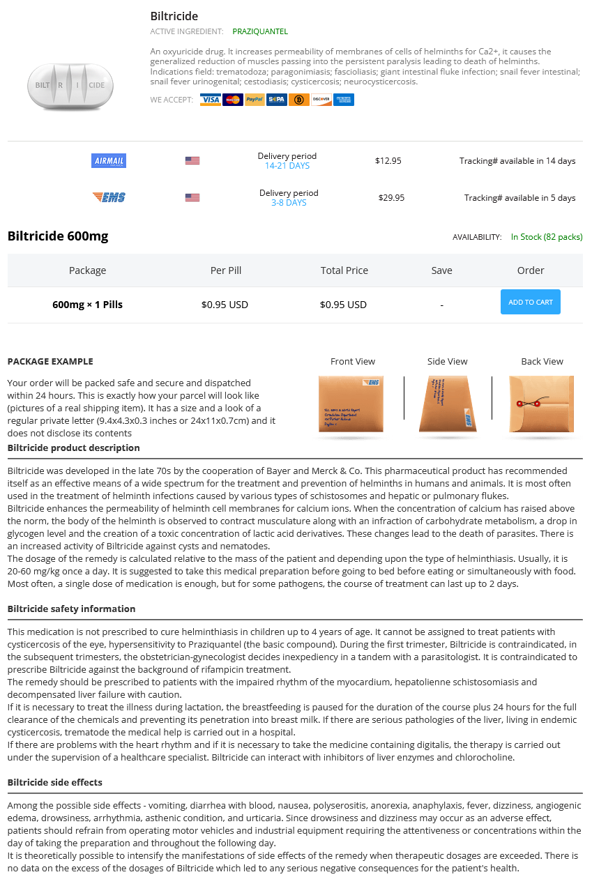
Biltricide
General Information about Biltricide
Biltricide is generally well-tolerated and has few side effects, which can include mild abdomen upset, dizziness, and headache. However, it should not be taken by individuals who're allergic to praziquantel or have liver illness, as it can worsen these circumstances. It is also not really helpful for use during being pregnant or whereas breastfeeding.
Cestodiasis, also referred to as tapeworm infection, is another situation that may be effectively handled with Biltricide. This type of an infection may be caused by a variety of tapeworms, which might infect the intestines or other organs, depending on the species. Biltricide helps to expel the tapeworms from the physique, stopping them from causing any further damage.
Biltricide can be used to deal with cysticercosis, a condition caused by the larval form of the pork tapeworm. This an infection occurs when the eggs of the tapeworm are ingested via contaminated meals or water. Biltricide helps to kill the larvae, preventing them from developing into grownup tapeworms.
Indications for using Biltricide include trematodiasis, a condition brought on by flukes; paragonimiasis, an an infection caused by lung flukes; and fascioliasis, an an infection attributable to liver flukes. It is also used to deal with infections brought on by the large intestinal fluke, a type of liver fluke found in Southeast Asia and the Pacific Islands.
Biltricide can additionally be effective in treating snail fever, also referred to as schistosomiasis, which is a sort of parasitic infection attributable to trematodes. This contains both intestinal and urinogenital types of the illness. The drug helps to eliminate the worms from the body, thereby relieving symptoms and preventing additional problems.
Biltricide, additionally known by its generic name praziquantel, is an oxyuricide drug used to treat varied forms of parasitic infections. It works by increasing the permeability of membranes of cells belonging to helminths, a kind of parasitic worm, for calcium ions. This leads to the generalized reduction of muscular tissues, in the end causing paralysis and dying of the helminths.
In conclusion, Biltricide is a strong oxyuricide drug that is used to deal with a broad range of parasitic infections. By increasing the permeability of cell membranes of helminths, it causes their paralysis and dying, leading to the elimination of the worms from the physique. This drug has a proven observe report of successfully treating numerous kinds of helminthiasis and is a generally prescribed treatment for individuals living in areas the place these infections are prevalent. However, it is important to seek the assistance of a healthcare professional earlier than using Biltricide to make sure it is the right remedy in your specific situation.
Biltricide is primarily used to treat infections attributable to several kinds of helminths, similar to trematodes, flukes, cestodes, and tapeworms. It can be efficient against sure forms of protozoa. The drug is available in the form of tablets and is usually administered orally.
One of probably the most serious conditions that can be treated with Biltricide is neurocysticercosis, a parasitic infection that impacts the central nervous system. This situation is caused by the ingestion of the eggs of the pork tapeworm, which then travel to the mind or spinal cord and might trigger critical neurological symptoms. Biltricide helps to kill the larvae and reduce the inflammation attributable to the an infection.
Mucus treatment 5th disease purchase biltricide 600mg on-line, the constituents of which are produced by goblet and epithelial cells, has a number of important protective functions and these are summarized in Table 2. These are glycoproteins with high proportions of carbohydrates-usually between 70 and 85% (w/w). The protein backbone consists of several thousand amino acid residues and contains regions with many oligosaccharide side chains and other regions without such side chains. The oligosaccharide-rich regions are resistant to proteases whereas the other regions are protease sensitive. The oligosaccharide-containing regions of the protein are rich in serine, threonine, and proline. This structure is maintained by repulsive forces operating between the negatively charged carbohydrate side chains and between the side chains and the protein backbone. The side chains also protect the protein from degradation by proteases and so help to preserve the integrity of the mucus gel. The side chains usually consist of between 2 and 12 residues from a limited range of sugars-usually galactose, fucose, N-acetylglucosamine, N-acetylgalactosamine, mannose, and sialic acids. The region of the side chain involved in linkage to the protein is known as the core, which is itself linked to a backbone consisting of a number of repeating disaccharide units containing galactose and N-acetylglucosamine. The terminal sugars are known as peripheral residues and often consist of sialic acid or sulfated sugars. The individual glycoprotein molecules are large (usually approximately 105106 Da) but in the mucus layer they are generally present as even larger molecules due to the formation of disulphide bonds that link the protein constituents of neighboring molecules. These polymers may be several micrometers in length and form a viscoelastic gel covering the mucosal surface. The mucus coating often has a monomolecular layer of lipids on its external surface that renders it hydrophobic. Each corneocyte is anucleated and contains a water-insoluble protein complex consisting mainly of a keratin microfibrillar matrix. The corneocytes are continually being shed into the environment taking any attached microbes with them. Bacteria are stained red using a universal oligonucleotide probe whereas epithelial cell nuclei are stained blue. Bacteria can be seen to be localized far away (approximately 30 µm) from the epithelium, either as dispersed cells or as microcolonies, in the outer regions of the mucus layer. The inner layer adheres firmly to the epithelium and contains a high concentration of mucin whereas the outer layer has a lower mucin content and is only loosely attached to the inner layer. In addition to mucins and lipids, the mucus coating also contains exfoliated cells, the contents of dead host cells, antibodies, and a range of antimicrobial compounds (listed in Appendix 1) produced by the mucosa. In the respiratory tract and cervix, the mucus coating is continually propelled along the mucosal surface by means of cilia-this is described in greater detail in Chapters 4 and 7. The antimicrobial peptides were detected by immunostaining with specific antibodies- staining (brown regions) was most dense in the uppermost layers of the epithelium. Most of the carbohydrates are linked to the protein at serine and threonine residues via an oxygen atom. A typical mucosal surface is covered with a mucus coating which often has a thin lipid layer on its outer surface. The mucins in the mucus coating interact with the membrane-bound mucins (glycocalyx) of the epithelial cells. The right panel is an expanded image of the corresponding boxed region in the left panel. Degradation of the mucus layer requires a number of enzymes acting in a specific order Although mucus plays an important role in host defense, a number of microbes are able to use mucins as a source of carbon, nitrogen, and energy. This is particularly important at those body sites where there may be few alternative nutrient sources, for example the urinary and respiratory tracts. However, the complexity of these polymers means that their complete breakdown cannot usually be achieved by a single microbial species. Degradation usually requires the production of a range of enzymes in a certain order because certain regions of the molecule only become accessible once others have been removed-this is more readily accomplished by microbial consortia rather than by individual species. Nevertheless, a limited number of microbes can achieve complete degradation of a mucin and these include the intestinal organisms Ruminococcus torques, Ruminococcus gnavus, a Bifidobacterium sp. The boxed regions in both (A) and (B) are shown at a higher magnification in the top left hand corner of each image. The sialic acid itself can be further degraded by acetylneuraminate pyruvate lyase to Nacetylmannosamine which can be used as a carbon and energy source by some bacteria. Sialidases (neuraminidases) Exoglycosidases Endoglycosidases Cleave sugars from side chains. Peptidases/proteases Cleave at non-glycosylated regions; degrade protein backbone after side chains have been removed. The range of enzymes needed to achieve complete degradation of a mucin is shown in Table 2. The ability to degrade mucin, entirely or partially, is found in microbes or microbial consortia inhabiting all mucosal sites and these will be described in appropriate sections of Chapters 4 to 9. The complete removal of mucus from a mucosal surface would leave the underlying epithelium vulnerable to microbial colonization and would have other harmful consequences. However, this does not appear to happen very often which means that mucus utilization by the indigenous microbiota must occur at the same rate as it is replaced by the host-another example of the balanced relationship that exists between the host and its indigenous microbes. The composition of the microbial communities that inhabit humans varies from site to site on the body.
Poor neurological state on admission and loss of consciousness at ictus have been associated with increased risk of early seizures symptoms carpal tunnel biltricide 600mg with visa. They commonly occur within the first 24 hours after ictus with 75% within 4 days in 686 Chapter 93: Epilepsy Associated with Subarachnoid Hemorrhage Table 93. Factors associated with poor short- and long-term outcome include age, the severity of neurological injury, and hemorrhage load. In case of equipoise of the two methods, endovascular coiling should be considered [2,3]. Before aneurysm obliteration, systolic blood pressure should be controlled (<160 mm Hg) to prevent rebleeding. Lumbar drainage can be used as an alternative and has been shown to reduce delayed cerebral ischemia but not to affect 6-month outcome [34]. When the aneurysm is surgically treated early after the bleeding, the outcome is more determined by medical complications [35], mechanisms resulting in secondary brain injury, including delayed cerebral ischemia, and seizures [21]. In case of confirmed vasospasm, other options include angioplasty with balloons or selective vasodilator drug infusions. Cerebral four-vessel three-dimensional rotational angiography is the gold standard for the detection and localization of ruptured aneurysms. Mortality rates decreased over the last decades to ~26% of hospitalized patients resulting in an increased number of survivors. However, the majority of the survivors have even in the long term cognitive deficits, decreased quality of life, and fatigue. This information has been used in outcome prediction models that showed an independent association with functional outcome and quality of life [15]. A more sophisticated approach calculating the time spent with seizures as cumulative seizure burden showed a linear correlation between this seizure burden and cognitive and functional outcomes [22]. These changes have been found to occur 2448 hours prior to changes seen with other diagnostic tools. Besides that, interictal and background findings such as sleep architecture, reactivity, and periodic epileptiform discharges were seen as relevant for prognostication [15]. No randomized controlled trials have been conducted to guide decisions on seizure prophylaxis or treatment. The use of phenytoin was furthermore associated with known side effects including impaired liver function, thrombocytopenia, rash, and StevensJohnson syndrome [10,47]. As previously discussed, there is some evidence that nonconvulsive seizures have the potential to cause additional injury in the acutely brain-injured patient. Treatment recommendations therefore derive from clinical centers and expert opinions. In patients with recurrent seizures, extended treatment for 46 weeks with re-evaluation thereafter is proposed [21]. Most authors agree that prolonged seizures are associated with brain damage and therefore secondary neuronal injury. One hypothesis is that seizure activity leads to additional brain injury in at-risk brain tissue through an excessive metabolic demand. The authors found a linear correlation between this seizure burden and both cognitive and functional outcomes [22]. The majority of these patients had only 2 seizures, which opens the discussion of a causal association [16]. Report of World Federation of Neurological Surgeons Committee on a Universal Subarachnoid Hemorrhage Grading Scale. Eugene Ramsay Cerebrovascular diseases are among the commonest causes of epilepsy and the most frequent causes of death and disability worldwide. Early seizures may not progress to epilepsy, but may result in increased morbidity and mortality. Based on the neurobiology and pathophysiology of cerebrovascular disease, there is little evidence to support the use of any specific anti-seizure medication when stroke is the cause of seizures. The best approach is stroke prevention, and, in lieu of that, design of future treatments should be aimed at reducing cell death and the cascade of events which may lead to development of epilepsy. There are limited data available regarding morbidity and mortality resulting from seizures after a stroke. However, at least one study, the Canadian Registry, showed a mortality rate at 30 days of 36. The presence of seizures may also lead to longer hospital stays and disability [1]. Individuals over 70 years of age account for twothirds of those affected with stroke and it is the leading cause of disability in adults. According to the World Health Organization, more than 50% of patients who had a stroke will have a residual deficit [8]. Overview of Cerebrovascular Disease Pathophysiology the initiating event for ischemic stroke is one of three pathophysiologic occurrences resulting in the interruption of oxygen and glucose delivery to brain cells [9]: (1) occlusion of a vessel by an embolus that develops at a distant site (2) thrombosis of an intracranial vessel (3) hypoperfusion of intracranial or extracranial arteries causing limited blood flow [1013]. Patients with hypercoagulable disorders are at risk for thrombosis leading to ischemic stroke and the risk may be unknown until the first presentation [14,15]. Once one of these initial events occurs, a cascade of actions occurs along two major pathways: the first pathway leads to cell death owing to the energy failure of the cell, and the second pathway consists of apoptosis or programmed cell death. Cell death can occur within 4 minutes if there is a total lack of oxygen and there is no collateral blood flow to the area of the infarction. Glutamate is also released upon depolarization and causes a rise in neuronal calcium influx.
Biltricide Dosage and Price
Biltricide 600mg
- 1 pills - $0.95
After delivery medications you cant drink alcohol with biltricide 600mg buy low price, mastitis, bacteremia, sepsis, meningitis, endometritis, and wound infections are possible. A number of predisposing factors have been identified and these include oral contraceptive use, pregnancy, diabetes mellitus, and broad spectrum antibiotic therapy. The condition usually responds to antifungal agents which can be administered either intravaginally (imidazoles- butoconazole, clotrimazole, or miconazole) or orally (fluconazole). The penetration of epithelial cells by hyphae results in the death of the host cell but there is also evidence that Can. Other important virulence factors include various hyphal proteins that inhibit phagocytosis and a large number of adhesins that enable both forms of the organism to adhere to the vaginal epithelium. Approximately 15 million preterm babies are born each year and this represents 11% of all live births worldwide. It is a major cause of perinatal mortality, serious neonatal morbidity, and childhood disability in both developed and developing countries. Microbial invasion of the uterus generates an inflammatory response involving the release of cytokines, chemokines, and prostaglandins, which induce the characteristic features of labor-uterine contractions and rupture of the fetal membranes. Predominant among these are organisms from the vaginal and/or cervical microbiotas such as Ureaplasma spp. This implies that the most common route of infection is by ascension through the vagina and cervix. Some of the organisms detected are generally regarded as being members of the oral and respiratory microbiotas and include Bergeyella spp. Common infections of the oral cavity such as gingivitis and periodontitis enable access of oral microbes to the bloodstream which can transport them to the placenta from where uterine invasion can take place. The disease can be diagnosed clinically on the basis of the following four symptoms: (1) the vaginal fluid has a pH greater than 4. Frequency of detection of various bacteria in the intrauterine microbiota of 349 pregnant women with intra-amniotic infection. The prevalence of the disease varies widely from country to country and within countries and is markedly affected by socioeconomic status, sexual activity and ethnicity. Photomicrograph of a vaginal sample showing a normal vaginal epithelial cell (the lower of the two cells) and another (the upper cell) with its exterior covered in bacteria giving it a roughened, stippled appearance known as a clue cell. The presence of clue cells is one of the diagnostic criteria of bacterial vaginosis. Because of diffusion limitations within the biofilm, a wide range of environments exist within it which enables the proliferation of different physiological types of microbes. Confocal laser scanning images of polymicrobial biofilms in vaginal samples obtained from two women with bacterial vaginosis (×400). Metabolomic studies have shown that the dysbiosis is accompanied by a change in the types of metabolites present in the vaginal ecosystem. Consequently, there is a decrease in the concentration of lactic acid, an increase in the concentrations of acetate, propionate, butyrate, and succinate, and an increase in amines such as putrescine, cadaverine, trimethylamine, and ethanolamine. Virulence factors of organisms associated with bacterial vaginosis and their possible role in the disease. It is a dysbiosis in which lactobacilli are usually absent and the microbiota is dominated by Strep. It is a serious condition with a high morbidity-20% of those affected become infertile and 20% develop chronic pelvic pain. Repeated douching can lead to vaginal dysbiosis Vaginal douching (the cleansing of the vagina with a liquid) is a practice carried out by many women for a variety of reasons including personal hygiene, aesthetic reasons, to prevent or treat an infection, to cleanse after menstruation or sex, and to prevent pregnancy. A variety of solutions have been used in douching including vinegar, various antiseptics, and detergents. The effects of douching on the vaginal microbiota depend on the nature of the solution used and how often it is practiced. However, the weight of evidence suggests that repeated douching results in dysbiosis with a decrease in the proportion of lactobacilli. Some forms of sexual behavior can result in vaginal and cervical dysbiosis A number of studies have shown that frequent sexual intercourse (more than one sex act per week), having multiple sex partners (three or more sex partners per year), frequent episodes of receptive oral sex (more than three per month), and the use of a spermicide result in dysbiosis of the vaginal microbiota. Women who have sex with women are also at greater risk of dysbiosis of the vaginal microbiota. The use of a copper intrauterine contraceptive device results in dysbiosis of both the vaginal and cervical microbiotas. The figure shows the proportions of women colonized by particular organisms before and after administration of the antibiotic. For example, a 10-day course of clarithromycin, a macrolide antibiotic, has been found to eliminate lactobacilli from the vagina and to increase the frequency of colonization by E. Proportion of women who develop vulvovaginal candidiasis within 7 weeks of the administration of various antibiotics. On the other hand, pivmecillinam (a narrow-spectrum penicillin that is active mainly against Gram-negative species) appears to have little effect on the cultivable microbiota of the vagina or cervix. The effects of other antibiotics on the cultivable microbiota of the vagina are summarized in Table 7. The presence of lactobacilli in the vagina has for many years been regarded (not necessarily correctly, for reasons discussed previously) as synonymous with vaginal health and, in view of the fact that many probiotics consist of lactobacilli, their use in the prevention and treatment of vaginal infections has been the subject of enormous interest for many years. Consequently, it was thought that maintaining high proportions of lactobacilli in the vagina by direct application of a probiotic should exert a beneficial effect.

ADDII BIOTECH is proud to announce that the company's manufacturing facility located at Baddii, Himachal Pradesh has recently been approved by Food & Drug Authority of Ghana.
Our Manufacturing capability includes a wide range of therapeutic products covering almost every segment.
ADDII BIOTECH given our strong emphasis on product quality and services.