
Cardura
General Information about Cardura
High blood stress, also identified as hypertension, is a standard situation that impacts tens of millions of individuals worldwide. It is sometimes called a silent killer as a result of it often has no warning signs or symptoms until severe problems arise. If left uncontrolled, high blood pressure can result in critical health issues such as coronary heart illness, stroke, and kidney failure. Cardura helps to lower and maintain a healthy blood strain level, thereby decreasing the risk of these dangerous circumstances.
Like any medicine, Cardura could trigger some unwanted effects, although not everyone experiences them. Common unwanted side effects embody dizziness, lightheadedness, nausea, and complications. These normally subside as the body adjusts to the treatment. In some cases, critical side effects similar to fainting, problem respiration, and chest pain could occur, and immediate medical attention should be sought if these occur.
BPH, on the other hand, is a non-cancerous enlargement of the prostate gland that may cause troublesome urinary signs in men. As men age, the prostate naturally grows in measurement, which might trigger pressure on the bladder and urethra, leading to issue in urination and other signs such as frequent urination, weak urine stream, and the feeling of incomplete bladder emptying. BPH mainly affects men over the age of fifty and might tremendously affect their quality of life. Cardura works by relaxing the muscles in the prostate and the bladder, making it easier for males to urinate.
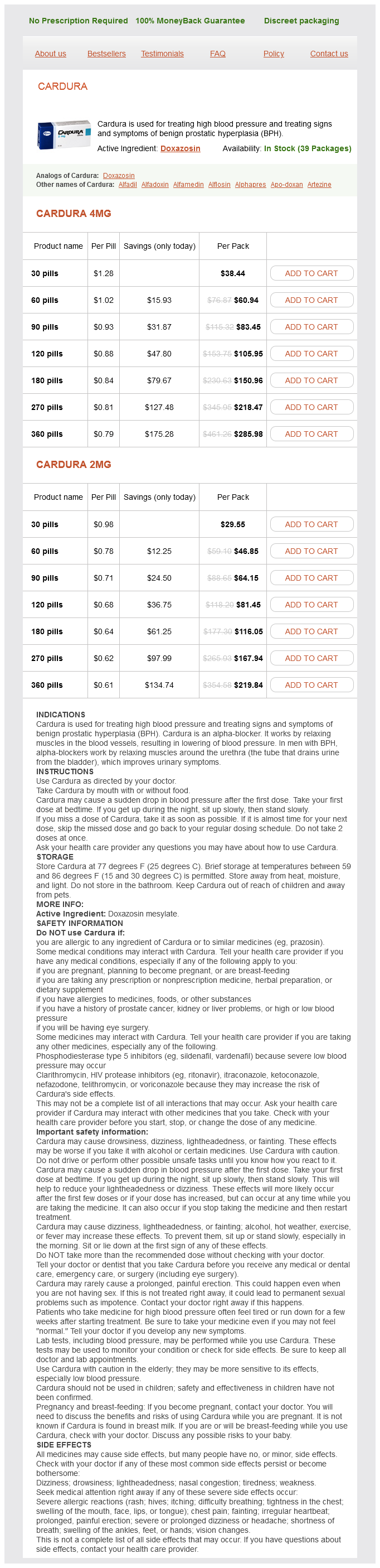
Cardura, also recognized by its generic name doxazosin, is a medication that's generally used for two major purposes: treating high blood pressure and managing signs of benign prostatic hyperplasia (BPH). This powerful drug belongs to a category of medications generally identified as alpha blockers, and has been confirmed to be an efficient remedy choice for patients suffering from these situations.
In conclusion, Cardura is a extremely efficient medicine for managing high blood pressure and urinary symptoms in men with BPH. It has been used for many years and has confirmed to be protected and well-tolerated by most patients. However, it is essential to observe the recommended dosage and to inform your doctor of any side effects or interactions with different medications. With correct use and monitoring, Cardura might help improve the quality of life for these dwelling with hypertension or BPH.
The major operate of Cardura is to relax and widen the blood vessels, which in turn lowers blood pressure and allows blood to circulate extra simply by way of the body. This lowers the risk of coronary heart attacks, strokes, and other cardiovascular problems. In addition, Cardura also works to enhance urinary signs in males with BPH by relaxing the muscular tissues in the prostate and bladder, making it simpler to urinate.
It is important to inform your physician about any other drugs you're taking earlier than beginning Cardura. It could interact with other drugs and cause potentially dangerous unwanted effects. It must also be used with warning in patients with liver or kidney disease, in addition to in those with low blood strain.
Cardura comes within the form of oral tablets and is normally taken once a day with or without meals. The dose could also be adjusted by a healthcare provider based mostly on the individual's response to the medicine. It is essential to take Cardura on the same time every day to maintain a gentle degree of the drug within the physique. It could take several weeks for the medication to indicate its full results, so it's important to not cease taking it with out consulting a physician.
The formation of central glutamatergic synapses involves similar developmental events but somewhat different specific players (McAllister hypertension 10 cardura 4 mg free shipping, 2007). Axodendritic contacts may be facilitated by cell adhesion molecules, but are also influenced by neuroligins and neurexins. Deficits in one or more of these complex signals may underlie psychiatric or neurodevelopmental disorders. The study of synapse formation has benefited from some truly novel imaging approaches over the years, including fluorescent imaging of axons in living animals over time (McCann et al. The novel "Brainbow" mouse transgenic lines were developed to visualize neuromuscular form, and their physiology can be extended to study neural circuits throughout the nervous system (Livet et al. Genomes of the Brainbow mice contain a transgenic insert consisting of randomly arranged, fluorescent protein genes separated by loxP sites and driven by neural/glialexpressing Thy1 promoter. After Cre recombination, the transgene undergoes excision events that position a different fluorescent protein gene adjacent to the promoter, resulting in its expression so that each neuron expresses one of a wide variety of possible fluorescent protein combinations. Some rearrangement occurs as the result of spontaneous activity during embryogenesis, but the majority of synaptic rearrangement happens after birth and as a result of sensory experience or motor activity. Crucial studies elucidating general principles of postnatal nervous system development were performed by Torsten Weisel and David Hubel who, along with Roger Sperry, shared the Nobel Prize in 1981 for their work on the development of the visual system. Blocking neural activity with tetrodotoxin prevents the segregation, indicating that neural activity must be important. Segregation appears to require synaptic stabilization of coordinately active cells. However, these cortical domains are still "plastic" and can change with experience. In classical studies, Hubel and Weisel closed one eyelid experimentally so that eye had no meaningful visual experience. When such monocular deprivation is started after birth, the "open-eye" columns expand as axons take over territory, while "closed eye" columns shrink. The process can be reversed by opening the previously shut eye and closing the open one, and again, the resulting axonal territories from each eye expand or contract to reflect the amount of activity in the respective eye. These synaptic rearrangements are not only dependent on activity, but are also dependent on experience, reflecting the quality of the visual experiences of the open eye. However, this remarkable plasticity does not remain forever; when monocular deprivation is initiated later in life, the cortical domains do not change and more connections appear fixed. Experience and synaptic plasticity in learning and memory are discussed further in Chapter 56. In a way, however, explaining the structure of the brain is only part of the story, as the brain is notorious for allowing us to learn throughout our lifetimes. There is a close relationship between the experience-dependent brain development that we just discussed and the learning and memory that go on throughout adulthood. Processes of development and learning probably are based on similar neural mechanisms, but occur at different times in the life of the organism and involve different brain areas. The chick/ quail transplantation model: Discovery of the isthmic organizer center. Retinal axon growth cones respond to EphB extracellular domains as inhibitory axon guidance cues. Graded activity of transcription factor Runx3 specifies the laminar termination pattern of sensory axons in the developing spinal cord. Prevention of the first occurrence of neural-tube defects by periconceptional vitamin supplementation. Interaction between Reelin and Notch signaling regulates neuronal migration in the cerebral cortex. Genes and signaling events that establish regional patterning of the mammalian forebrain. Neuronal specification in the spinal cord: Inductive signals and transcriptional codes. Transgenic strategies for combinatorial expression of fluorescent proteins in the nervous system. In vivo imaging of presynaptic terminals and postsynaptic sites in the mouse submandibular ganglion. A role for Gbx2 in repression of Otx2 and positioning the mid/hindbrain organizer. Involvement of bone morphogenetic protein-4 and bone morphogenetic protein-7 in the differentiation of the adrenergic phenotype in developing sympathetic neurons. Floor plate and motor neuron induction by different concentrations of the amino-terminal cleavage product of sonic hedgehog autoproteolysis. Bone morphogenetic proteins are required in vivo for the generation of sympathetic neurons. Effect of a notochordal implant on the early morphogenesis of the neural tube and neuroblasts: Histometrical and histological results. Growth factors are proteins that regulate many aspects of cellular function, including survival, proliferation, migration and differentiation. In non-neuronal cells growth factors stimulate proliferation, but mature neurons are postmitotic and cannot re-enter the cell cycle. Consequently, when considered in the context of the nervous system, growth factors are frequently referred to as neurotrophic factors. These factors are critical for proper development of the nervous system from the earliest embryonic stages.
The lipid electron-transport molecule ubiquinone (Q9 heart attack kiss buy cardura 1 mg lowest price, Q10) is the main site of free radical leaks from mitochondria via the ubiquinone cycle. Mitochondria appear to be an important and perhaps dominant source of free radicals in brain tissue subjected to ischemiareperfusion. Elevated concentrations of intracellular Ca2 and Na, a consequence of energy failure and excitotoxic glutamate receptor stimulation, can be expected to inhibit complex I as well as overproduction of superoxide anion. The resultant oxidative stress may lead to further inhibition of mitochondrial respiratory components, promoting further free radical production in a vicious, feed-forward cycle. Although xanthine oxidase has been implicated in animal models of ischemia, its role in human stroke is less clear (see also in Ch. In particular, cyclooxygenase isoforms and 5-lipoxygenase contain heme iron and may generate a low concentration of superoxide anion constitutively. Catalase and glutathione peroxidase provide two important cellular systems for eliminating H2O2. It can be irreversibly inactivated by oxidation and it demonstrates decreased activity after ischemiareperfusion. Catalase is more abundant in astrocytes than in neurons and in white matter than in gray matter, but it can be induced in neurons by neurotrophins. There is substantially less catalase activity in brain than in other tissues, such as liver. In addition, it has a cytosolic isoform and an isoform with a mitochondrial targeting sequence. Several other proteins, including glutathione S-transferase, may contribute minor peroxidase activity. Because humans, in contrast to most other animals, are unable to synthesize vitamin C, this important antioxidant must be supplied entirely from dietary intake. Other proteins, such as thioredoxin and metallothionein, may also contribute to some extent to the cellular antioxidant pool. Free radicals also contribute to apoptosis at several points in the apoptotic cascade, serving as initiators, early signals and possibly late effectors of apoptotic neuronal death (Nakka et al. It may be this interaction of injury mechanisms (including excitotoxicity, ischemic apoptosis, oxidative injury, inflammation and impaired metabolism) that, in part, makes the brain so vulnerable to ischemic damage. A conceptual framework, the neurovascular unit, comprises neurons, the microvessels that supply them, and their supporting cells. Cerebral microvessels consist of the endothelium (which forms the bloodbrain barrier), the basal lamina matrix, and the end-feet of astrocytes. Communication has been shown to occur between neurons and microvessels through astrocytes. As a result of the breakdown of the neurovascular unit after ischemia, the tissue content of water in the affected area of the brain may increase, leading to swelling or edema of the ischemic region. Some of the increased tissue water is intracellular, reflecting impaired cellular ion homeostasis due to energy failure, as well as the action of glutamate, which triggers excess cation influx in neurons and glia. Damage to microvascular endothelial cells can lead to vasospasm and can promote adhesion of platelets and leukocytes, leading to vessel plugging. Reperfusion injury is an inflammatory process modulated by the complex signaling mechanisms of the immune system. With reperfusion, additional neutrophils, platelets, and other inflammatory cells from the peripheral circulation rapidly accumulate within the brain tissue in response to the signals of chemotactic agents (Hall et al. There is swelling of endothelial cells and interstitial edema, both of which lead to increased hydrostatic pressures within the microcirculation and narrowed capillary lumina. There is loss of adrenergic control of vascular tone and loss of production of vasodilating agents such as nitric oxide from the endothelial cells. Leukocytes and platelets plug the postcapillary venules, further impairing capillary flow (Mehta et al. Agents that protect endothelial cells, such as antioxidants, can help preserve neurovascular unit integrity following ischemiareperfusion (Hall et al. An extensive infarct involves the dorsolateral and lateral portions of neocortex and the entire striatum. Managing patients with malignant cerebral infarction remains one of the foremost challenges in medicine. Despite the clinical significance of cerebral edema, the mechanism of brain water transport and edema formation remains poorly understood. The release of osmotically active substances (arachidonic acid, electrolytes, lactic acid) from necrotic brain tissue causes cerebral edema. This is aggravated by vascular injury and leakage of proteins into the interstitial space. Various cytokines peak in the serum within the first 24 hours of an acute stroke, and are thought to initiate the cascade of tissue damage. At the site of ischemia itself, activated leukocytes release free radicals and toxins causing further destruction. By three to four days, interstitial fluid accumulates in the infarct and around it. Finally, because tissue edema in the rigid intracranial cavity can restrict blood flow, secondary ischemia can ensue. However, they are induced rapidly in response to cerebral ischemia, in which case they exert diverse actions. Some elements that may be involved in this inflammatory response include macrophages, endothelial cells, neutrophils, lymphocytes and platelets, along with noncellular elements, such as the complement system, the blood Significance of aquaporins in brain edema Recently, aquaporins have been implicated in edema formation after ischemia (Zador et al. Aquaporins are small, homotetrameric membrane channels that mediate rapid water flux through cell membranes (see Aquaporins in Ch. Under these conditions, microvascular perfusion also might be impaired (Lucas et al. The presence of damaged cells and debris causes ramified resting microglia to transform into rounded migratory macrophages (de Groot & Rauen 2007).
Cardura Dosage and Price
Cardura 4mg
- 30 pills - $38.44
- 60 pills - $60.94
- 90 pills - $83.45
- 120 pills - $105.95
- 180 pills - $150.96
- 270 pills - $218.47
- 360 pills - $285.98
Cardura 2mg
- 30 pills - $29.55
- 60 pills - $46.85
- 90 pills - $64.15
- 120 pills - $81.45
- 180 pills - $116.05
- 270 pills - $167.94
- 360 pills - $219.84
Reperfusion therapies for acute ischemic stroke: Current pharmacological and mechanical approaches arteria gastrica sinistra cardura 2 mg purchase. Molecular mechanisms of apoptosis in cerebral ischemia: Multiple neuroprotective opportunities. Matrix metalloproteinase-9 as a marker for acute ischemic stroke: A systematic review. Systemic fatty acid responses to transient focal cerebral ischemia: Influence of neuroprotectant therapy with human albumin. Early changes in blood brain barrier permeability to small molecules after transient cerebral ischemia. Synergistic effects of a combination of low-dose basic fibroblast growth factor and citicoline after temporary experimental focal ischemia. Neuroprotective efficacy of combination therapy with two different antioxidants in rats subjected to transient focal ischemia. The oxidation of arachidonic acid by the cyclooxygenase activity of purified prostaglandin H synthase: Spin trapping of a carbon- centered free radical intermediate. Mice overexpressing extracellular superoxide dismutase have increased resistance to focal cerebral ischemia. Blockade of N-methyl-D-aspartate receptors may protect against ischemic damage in the brain. The importance of the omega-6/omega-3 fatty acid ratio in cardiovascular disease and other chronic diseases. Safety of mechanical thrombectomy and intravenous tissue plasminogen activator in acute ischemic stroke. Reactive oxygen radicals and pathogenesis of neuronal death after cerebral ischemia. Treatment of embolic stroke in rats with bortezomib and recombinant human tissue plasminogen activator. Temporal profile of ischemic tissue damage, neutrophil response, and vascular plugging following permanent and transient (2H) middle cerebral artery occlusion in the rat. A new mouse model for temporal- and tissue-specific control of extracellular superoxide dismutase. Excitable membranes have a phospholipid composition that differs from that of other membranes, a property assumed to be related to their highly specialized functions. The understanding of excitable membrane organization has conceptually evolved from the lipid bilayer with embedded proteins to a highly dynamic, heterogeneous patchwork of microdomains that contain ion channels, receptors, transporters and other proteins. Cellular membranes in the nervous system were divided in the past into relatively more fluid membranes. Several phospholipid pools in neurons, glia and endothelial cells of the cerebrovasculature are now recognized as reservoirs of lipid messengers. In addition, there are lipid rafts, which are cholesterol- and sphingolipid-rich microdomains in specific cellular compartments of the nervous system, including dendrites, where they are associated with specific postsynaptic proteins. Interestingly, there are subtypes of lipid rafts, which vary by their resistance to detergent extraction, their density and their raft marker proteins, Thy-1 and caveolin. Moreover, the normal density of synapses and dendritic spines seems to depend on lipid rafts, since changes in cholesterol availability modify rafts and in turn the properties of synapses and spines. Several lipid mediators generated in response to such stimuli regulate and interact with many other signaling cascades, contributing to the development, differentiation, function, protection and repair of the cells of the nervous system (Shimizu, 2009). They may be reincorporated into membrane lipids or further metabolized to biologically active derivatives. The remaining lysophospholipid can be either re-esterified and reincorporated into a membrane phospholipid or further metabolized. This chapter surveys the neurochemistry of lipid messengers, as well as the mechanisms by which bioactive lipids accumulate upon stimulation in response to injury, cerebral ischemia, seizures, neurotrauma or neurodegenerative diseases, and their significance in pathophysiology. However, these bioactive lipids have additional neurobiological actions in ion channel functions, receptors, neurotransmitter release, synaptic plasticity and neuronal gene expression. Bioactive lipids may be considered dual messengers: they modulate cell functions as messengers and they become part of the response of the nervous tissue to injury, broadly referred to as the inflammatory response. Some molecular species of phospholipids in excitable membranes are reservoirs of bioactive lipid mediators that act as messengers Signals, such as those resulting from neurotransmitter receptor occupancy, trigger the release of phospholipid moieties via the activation of phospholipases. The inhibitory effects of this antagonist on glutamate release could account in part for its neuroprotection in ischemiareperfusion. Synapses are intimately surrounded by astrocytes, which express glutamate transporters that remove the excitatory neurotransmitter from the vicinity of the synaptic cleft. Astrocytes also respond to prostaglandins by releasing glutamate through a Ca2-dependent mechanism, although the quantitative importance and therefore functional relevance of astrocyte exocytosis of glutamate and other astrocyte derived "gliotransmitters" has come into question (Hamilton & Attwell, 2010). Other neuronal correlates of the inflammatory response include signaling by cytokines, nitric oxide and various growth factors. Activation of arriving inflammatory cells also plays a role in initial defenses against injury, removal of cellular debris and the longer-term repair/wound healing of the nervous system. Several lipid messengers are released from these cells and may participate in beneficial actions. Much remains to be learned in this area of integration of our knowledge of the inflammatory response and neuroimmune/repair signaling. Identification of lipids with biological activity has progressed remarkably over the last decade. While the inositol phosphates have been known for some time to play fundamental roles in cell biology (see Ch. Each member of the 15 groups is also assigned to one of five more general categories called "types.

ADDII BIOTECH is proud to announce that the company's manufacturing facility located at Baddii, Himachal Pradesh has recently been approved by Food & Drug Authority of Ghana.
Our Manufacturing capability includes a wide range of therapeutic products covering almost every segment.
ADDII BIOTECH given our strong emphasis on product quality and services.