
Careprost
General Information about Careprost
One of the explanations for Careprost's reputation is its ease of use. Unlike other medications used for these situations, Careprost is on the market within the form of eye drops, making it simpler for patients to administer themselves. It also has a comparatively low threat of unwanted effects, with the commonest ones being delicate eye irritation or a darkening of the iris and eyelashes. However, these unwanted facet effects usually are not permanent and can be simply managed.
Careprost is a medication that belongs to a bunch of medicine referred to as prostaglandin analogs. It works by decreasing the stress in the eye by increasing the outflow of fluid from the attention. This, in turn, helps to decrease the danger of injury to the optic nerve. Careprost is usually used once per day in the affected eye(s) and has proven to be highly effective in reducing the strain in the eye and preventing additional injury.
Careprost is a drugs that has been gaining recognition lately for its ability to treat two common eye situations - ocular hypertension and open-angle glaucoma. Both of those situations contain a rise in strain within the eye, which may lead to critical imaginative and prescient problems if left untreated. Careprost has proven to be a highly efficient remedy for these circumstances, making it a best choice for a lot of sufferers and docs alike.
However, it's important to notice that Careprost should solely be used underneath the supervision of a physician. They will evaluate your eye health and determine if this medication is suitable for you. They will also monitor your progress and any potential unwanted side effects.
In conclusion, Careprost is a extremely effective and affordable medication for treating ocular hypertension and open-angle glaucoma. Its ease of use, low danger of unwanted aspect effects, and additional beauty advantages make it a most well-liked choice for each patients and medical doctors. If you're experiencing increased stress in your eyes, it is crucial to hunt medical consideration and discuss the potential for utilizing Careprost as a treatment choice. Remember, taking care of your eyes is vital for maintaining good imaginative and prescient and general well being.
Careprost has additionally confirmed to be a cost-effective remedy choice for ocular hypertension and open-angle glaucoma. It is considerably more affordable than different drugs used for these circumstances, making it accessible to a wider vary of patients. Additionally, since it is usually a life-long treatment, the fee savings can add up over time for sufferers.
In addition to its effectiveness in treating ocular hypertension and open-angle glaucoma, Careprost additionally has different beauty advantages. It has been found to promote eyelash growth and thickness, making it a preferred choice for these seeking to improve the looks of their eyelashes. This is due to the active ingredient in Careprost, bimatoprost, which also stimulates the expansion of hair follicles.
First and foremost, it is necessary to perceive what ocular hypertension and open-angle glaucoma are and the way they'll have an effect on our vision. Ocular hypertension is a situation during which the strain inside the eye is elevated, but there are no signs or indicators of injury to the optic nerve. It is commonly a precursor to open-angle glaucoma, which is a extra severe condition the place the optic nerve turns into broken as a result of prolonged excessive stress within the eye. If left untreated, open-angle glaucoma can result in everlasting imaginative and prescient loss.
Nuclear receptors are responsible for the action of the thyroid hormone tetraiodothyronine (T3) administering medications 7th edition ebook cheap careprost 3 ml online. T3 binding to the nonhistone receptor proteins stimulates transcription at the 12. Amine, polypeptide, and protein hormones (either growth promoting or inhibiting) initiate their action by binding to plasma membrane receptors on the cell surface. Monomeric G-proteins anchored to the inner cytoplasmic membrane also participate in the normal cellular functions when activated by external stimuli. Some of these are proto-oncogenes and, when mutated, become oncogenes that promote cancer. The first type contains an extracellular receptor domain and an intracellular domain with tyrosine kinase activity. Nonreceptor tyrosine kinases are found in the cytosol and are involved in the signal transduction pathways of normal cellular processes. Specific inhibitions at the receptor sites using monoclonal antibodies or tyrosine kinase inhibitors are therapeutic targets used in the management of some cancers. Endocrine topics not discussed in Chapters 28 through 32 that are covered elsewhere in the text are as follows: gastrointestinal hormones, Chapter 11; eicosanoids, Chapter 16; pancreatic hormones, Chapter 20; parathyroid hormone and vitamin D, Chapter 35; reninngiotensin system and antidiuretic hormone, Chapters 30 and 37. Natriuretic peptides initiate their action of vasodilation and natriuresis by binding to receptor-guanylyl cyclases. Nitric oxide initiates its action of smooth muscle relaxation by binding to cytosolic guanylyl cyclase. The supplemental reading list provides sources that describe the various pathways. All of these amines except the thyroid hormones (Chapter 31) are decarboxylated products that are synthesized both within and outwith the nervous system. Within the nervous system, they are important neurotransmitters; outside the nervous system, the cells that produce them are modified postsynaptic neurons. Regardless of where they are released, hormonal amines exert their effects through specific receptor sites located in various parts of the body. All of these amines, except T3, exert rapid systemic effects that usually involve smooth-muscle activity. Because the amines are hydrophilic, their receptors are located on the outer surface of target cells, and most if not all of their effects are mediated by intracellular mediators (discussed later). The survival of multicellular organisms depends on the integration and coordination of differentiated cell functions and the ability to react appropriately to internal and external influences that threaten to disrupt homeostatic conditions. These requirements are fulfilled by a form of intercellular communication in which chemical signals (messengers) released by one cell evoke a receptor-mediated response in another. Neurotransmitters convey signals from one neuron to another or from a neuron to an effector cell, travel very short distances to reach their target sites, and function within the specialized regions of synapses and junctions. Hormones are usually defined as messengers that are transported by the blood to distal target cells. Because they are released into the interstitial space and then into the blood, they are called endocrine (ductless; "secreted within") secretions to distinguish them from those that are released into the external environment ("exocrine" or ductal secretions). Hormones and neurotransmitters may have evolved from the same or similar ancestral prototypes in unicellular organisms. Thus, the structure of hormones and neurotransmitters and their functions as chemical messengers may have been highly conserved during evolution. The fact that many prototypical messengers in unicellular organisms predate the appearance of nerve cells implies that neurotransmitters may be a specialized form of hormone. More than 40 hormones have been identified, containing from three to over 200 amino acid residues, and they are discussed in various parts of the text. All hormonal peptides and many hormonal proteins are synthesized as part of a larger molecule (preprohormone) that contains a leader sequence (signal peptide) at its amino terminal end. The leader sequence is removed as the nascent precursor enters the lumen of the endoplasmic reticulum, and the resultant prohormone undergoes posttranslational processing after being packaged into secretory granules by the Golgi complex. Post-translational processing involves proteolytic cleavage at specific sites, usually paired basic amino acid residues (Lysrg, Argys, Argrg, Lysys), by endopeptidases within the secretory granule. In the synthesis of insulin, for example, removal of the 23-amino acid leader sequence of preproinsulin results in proinsulin, which has two pairs of basic residues (Lysrg and Argrg) that are cleaved 2. They have the ability to synthesize and release peptide hormones and, as their name implies, take up amine precursors. Amine hormones and some agents of the second type have counterparts in the nervous system that function as neurotransmitters. Likewise, the endogenous opiates arise from site-specific cleavages of their respective prohormones (Chapter 29). When the cell is stimulated to secrete, all major fragments of the prohormone, active and inactive, are released by calciumdependent exocytosis. Although several peptide hormones have multiple anatomical sites of synthesis, there is usually only one major source of the circulating hormone. The presence of the same hormone in ancillary sites may indicate that it functions as a local hormone at those sites. Other "brainut" peptides (Chapter 29) also illustrate this point, although they are not released into the general circulation in significant amounts. Several families of peptide hormones have been described, including the opiomelanocortin family (endorphins, adrenocorticotropic hormone, melanocytestimulating hormone), the somatomammotropin family (growth hormone, prolactin, human placental lactogen), the glycoprotein hormones (thyroid-stimulating hormone, luteinizing hormone, follicle-stimulating hormone, human chorionic gonadotropin), the insulin family (insulin, insulin-like growth factors, somatomedins, relaxin), and the secretin family (secretin, glucagon, glicentin, vasoactive intestinal peptide). Thus, members of the secretin family regulate secretory activity in target cells, while those of the insulin family promote cell growth.
Lipochrome/lipofuscin: Wear and tear pigment seen in old age medicine cat herbs buy cheap careprost 3 ml online, severe malnutrition, and cancer cachexia. Pigments are colored substances, which are either normal constituents of cells. Hemosiderin It is a hemoglobin-derived, golden yellow-to-brown, granular or crystalline pigment and is one of the major storage forms of iron. Causes Local or systemic excess of iron cause hemosiderin to accumulate within cells. H&E showHemochromatosis: Severe accumulation of iron is associated with damage to liver, heart, ing macrophage containing and pancreas. The triad of cirrhosis of liver, diabetes mellitus (due to pancreatic damage) coarse, golden granular pigment within the cytoplasm; and brown pigmentation of skin constitute bronze diabetes. This pigment is gives blue black color to hemosiderin pigment seen in chronic malaria and in mismatched blood transfusions. It is non-iron containing pigment Lipofuscin: Important indiderived from hemoglobin. Commonly used histochemistry (special stains) in histopathology are listed in Table 1. Lipid Red Brown black Extracellular collagen Collagen, smooth muscle Cross striation of skeletal muscles, glial filaments, fibrin Elastic fibers Red Collagen-blue, smooth muscle-red Dark blue Black Contd. Causes Aging is multifactorial and is affected by genetic factors and environmental factors. They cause progressive accumulation of sublethal injury over the years at cellular and molecular level. Mechanism of Cellular Aging Decreased Cellular Replication Most normal cells have a limited capacity for replication. After about 60 to 70 cell divisions, all cells become arrested in a terminally nondividing state, known as senescence. The following mechanisms may be responsible for progressive senescence of cells and decreased cellular replication in aging. Telomeres ensure the complete copying of chromosomal ends during the S-phase of the cell cycle. When telomeres are sufficiently shortened, cells stop dividing leading to a terminally nondividing state. Telomere shortening may be one of the mechanisms responsible for decreased cellular replication. Telomerase Telomerase is an enzyme that regenerates and maintains telomere length. In cancers, the telomerase may be reactivated in tumor cells resulting in maintenance of length of telomeres. Defective Repair Mechanism Many protective repair responses counterbalance the metabolic damage in cells. Thus, aging can be delayed by either by reducing the metabolic damage or by increasing the repair response to that damage. Factors that Increases Longevity Caloric Restriction Calorie restriction prolongs lifespan and this longevity appears to be mediated by a family of proteins known as sirtuin. Actions of Sirtuins:Sirtuins promotes the expression of many genes which increase longevity. The proteins products of these genes increase metabolic activity, reduce apoptosis, stimulate protein folding, and inhibit the damaging effects of oxygen free radicals. Growth Factor Signaling Growth factors, such as insulin-like growth factor trigger the insulin receptor pathway. This results in activation of transcription factors which activate genes that reduce longevity. Inflammation: Complex local response of the living vascularized tissues to injury. Lasts for hours or a few days Neutrophils (also called polymorphonuclear leukocytes) Exudation of fluid and plasma proteins (edema) and the emigration of leukocytes Usually mild and self-limited and can progress to a chronic phase Prominent Chronic inflammation May follow acute inflammation or be slow in onset (days) Longer duration; may be months Lymphocytes, monocytes/macrophages and sometimes plasma cells Inflammatory cells associated with the proliferation of blood vessels, tissue destruction and fibroblast proliferation Usually severe and progressive with fibrosis and scar formation Less prominent Q. Inflammation and the accompanying repair process is a beneficial host response in most instances, but can sometimes be harmful. Injury/damage to tissue and fibrosis Signs: Local and systemic Cardinal Signs of InflammationThe four cardinal signs of inflammation as mentioned by Celsus are listed in Table 2. Celsus described the first 4 cardinal signs of inflammation namely: Rubor, calor, tumor and dolor. Explain the sequential vascular changes/reactions of blood vessels/ hemodynamic changes in acute inflammation. Normal hydrostatic pressure in the capillary bed:About 32 mm Hg at the arterial end12 mm Hg at the venous end. Normal mean colloid osmotic pressure of tissues is about 25 mm Hg and is equal to the mean capillary hydrostatic pressure. Formation of exudate in inflammation Changes in Vascular Flow and Caliber Vasodilatation in acute inflammation isVasodilatation: It is the earliest feature of acute inflammation; sometimes it follows a responsible for the one transient constriction of arterioles. Increased Vascular Permeability (Vascular Leakage) Exudation: It is defined as the process of escape of fluid, proteins and circulating blood cells Increased vascular permeability: Hallmark of from the vessels into the interstitial tissue or body cavities. Escape of a protein-rich fluid causes edema and is one of the cardinal signs of inflammation. Mechanism of Increased Vascular Permeability Several mechanisms can cause increased vascular permeability: 1. Increased vascular permeability causes one of the cardinal signs of inflammation namely tumor (edema). Leukocyte-mediated vascular injury: the leukocyte (mainly neutrophils) which adheres to the endothelium during inflammation may themselves injure the endothelial cells. Increased transcytosis: Process of transport of fluids and proteins through the channels called vesiculovacuolar organelle is increased in number.
Careprost Dosage and Price
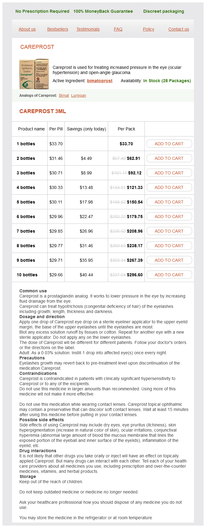
Careprost 3ml
- 1 bottles - $33.70
- 2 bottles - $62.91
- 3 bottles - $92.12
- 4 bottles - $121.33
- 5 bottles - $150.54
- 6 bottles - $179.75
- 7 bottles - $208.96
- 8 bottles - $238.17
- 9 bottles - $267.39
- 10 bottles - $296.60
Diphtheria is effectively prevented by immunization with toxoid (inactivated toxin) preparations symptoms stomach ulcer order 3 ml careprost overnight delivery. Thus, patients infected with Vibrio cholerae secrete enormous quantities (up to 20 L/d) of water and electrolytes. Without adequate and prompt replacement, death can ensue owing to dehydration and electrolyte imbalance. A group of plant lectins, such as abrin, ricin, and modeccin, are highly toxic to eukaryotic cells. Like most plant and bacterial toxic proteins, ricin contains two polypeptide chains with two different but complementary functions. The A chain possesses enzymatic activity and is responsible for toxicity, and the B chain, which is a lectin, binds to galactose-containing glycoproteins or glycolipids on the cell surface. On binding of the ricin molecule to the carbohydrate receptors of the cell surface via the B chain, the A chain enters the cytoplasm, presumably by receptor-mediated endocytosis, where it inhibits protein synthesis by irreversible inactivation of the 60S ribosomal subunit. Toxins have been used to develop highly selective cytotoxic agents targeted against specific cells. For example, ricin A has been coupled to agents that selectively bind to cell-surface membrane components. The selective agents may be monoclonal antibodies (Chapter 33), hormones, or other cell-surface ligands. These conjugates act as selective cytotoxic agents and may have potential therapeutic applications. Potential clinical application for diphtheria toxin may be impossible because of the prevalence of the diphtheria antitoxin in human populations. Collagen Biosynthesis and Its Disorders Collagen occurs in several genetically distinct forms and is the most abundant body protein; most of the body 442 Essentials of Medical Biochemistry scaffolding is composed of collagen (Chapter 11). It is a fibrillar protein but also exists in a nonfibrillar form in the basement membrane. The basic structural unit of collagen, tropocollagen, consists of three polypeptide chains. Some of the amino acid residues at the X and Y positions are proline, hydroxyproline, lysine, hydroxylysine, and alanine. Collagen, which is a glycoprotein, contains only two types of carbohydrate residues: glucose and galactose linked in O-glycosidic bonds to hydroxylysyl residues. In collagen, each polypeptide chain is coiled into a special type of a rigid, kinked, left-handed helix, with about three amino acid residues per turn. The three helical polypeptides, in turn, are wrapped around each other to form a right-handed triple-stranded superhelix that is stabilized by hydrogen bonding. The collagen molecules associate in an ordered quarter-staggered array to give rise to microfibrils, which, in turn, combine to give fibrils. Covalent cross-linkages occur at various levels of collagen fiber organization and provide great mechanical strength. Hydroxylation of selected prolyl and lysyl residues; Glycosylation of certain hydroxylysyl residues; Folding of procollagen polypeptides into a triple helix; Conversion of procollagen to collagen; Self-assembly into fibrils; Oxidative deamination of -amino groups of strategically located lysyl and hydroxylysyl residues to provide reactive aldehydes. These form cross-linkages between polypeptide chains of the same molecule as well as between the adjacent molecules that gives strength and stability to the fibrils. The first three processes take place inside the cell, whereas the last three are extracellular modifications. Collagen disorders can result from primary defects in the structure of procollagen or collagen or from secondary changes that affect collagen metabolism. Ehlersanlos syndrome and osteogenesis imperfecta are examples of inherited primary collagen diseases; scurvy and various fibrotic processes. Aberration in the control mechanisms that alter the balance between synthesis and degradation. This imbalance can lead either to excessive deposition or to depletion of collagen. Intracellular processes include hydroxylation of prolyly and lysyl residues, glycosylation of certain hydroxylysyl residues with glucose and/or galactose, and formation of procollagen triple helixes. Removal of terminal peptides by aminopeptidase and carboxypeptidase, formation of collagen fibrils, and extensive cross-link formation by lysyl oxidase occur in the extracellular matrix. Abnormalities in the packing of collagen molecules into a fiber or in the interaction of collagen fibers with other extracellular components of the connective tissue. These enzymes are located within the cisternae or rough endoplasmic reticulum; as the procollagen chains enter this compartment, the hydroxylations begin. All three enzymes have the same cofactor requirements: ferrous iron, -ketoglutarate, molecular oxygen, and ascorbate (vitamin C). The reducing equivalents required for the hydroxylation reaction are provided by the decarboxylation of equimolar amounts of -ketoglutarate to succinate and carbon dioxide: O C H2C these reactions are catalyzed by two specific enzymes: hydroxylysyl galactosyltransferase and galactosylhydroxylysyl glucosyltransferase. The substrate has to be in the nonhelical conformation, and the glycosylation ceases when the collagen propeptides fold into a triple helix. Thus, both hydroxylation and glycosylation must occur before triple-helix formation, which is an intracellular process. Deficiency of galactosylhydroxylysyl glucosyltransferase has been seen in the members of kindred with a dominantly inherited disease known as epidermolysis bullosa simplex. However, these proteins lack the proper modifications and therefore cannot be used for assembly of collagen fibrils. Thus, the role of ascorbate in the hydroxylation of collagen is one of its major physiological roles (see also Chapter 36).

ADDII BIOTECH is proud to announce that the company's manufacturing facility located at Baddii, Himachal Pradesh has recently been approved by Food & Drug Authority of Ghana.
Our Manufacturing capability includes a wide range of therapeutic products covering almost every segment.
ADDII BIOTECH given our strong emphasis on product quality and services.