
Chloramphenicol
General Information about Chloramphenicol
Chloramphenicol should not be utilized in sufferers with a historical past of blood issues, liver illness, or kidney issues. It can additionally be important to tell your doctor of any medications you are presently taking, together with over-the-counter medication and herbal dietary supplements, as they may interact with chloramphenicol.
Chloramphenicol works by stopping the expansion of micro organism, ultimately killing them. It does this by binding to bacterial ribosomes, which are responsible for producing proteins required for bacterial development and copy. By inhibiting the formation of those proteins, chloramphenicol halts the growth and spread of micro organism, allowing the body’s immune system to struggle off the infection.
Chloramphenicol is a broad-spectrum antibiotic that was first found in 1947. It is a naturally occurring compound produced by Streptomyces venezuelae, a soil bacterium. This antibiotic is widely available within the type of eye drops, ointments, capsules, and injections.
In rare instances, chloramphenicol could cause a critical situation referred to as aplastic anemia, where the bone marrow stops producing enough new blood cells. This condition could be life-threatening and requires instant medical consideration.
As with any treatment, chloramphenicol has potential unwanted effects. The most typical unwanted facet effects reported by sufferers include bone marrow suppression, which might trigger a decrease within the production of red and white blood cells and platelets. This can lead to an increased risk of infections, anemia, and bleeding issues. Other unwanted effects might embody nausea, vomiting, diarrhea, and skin rashes.
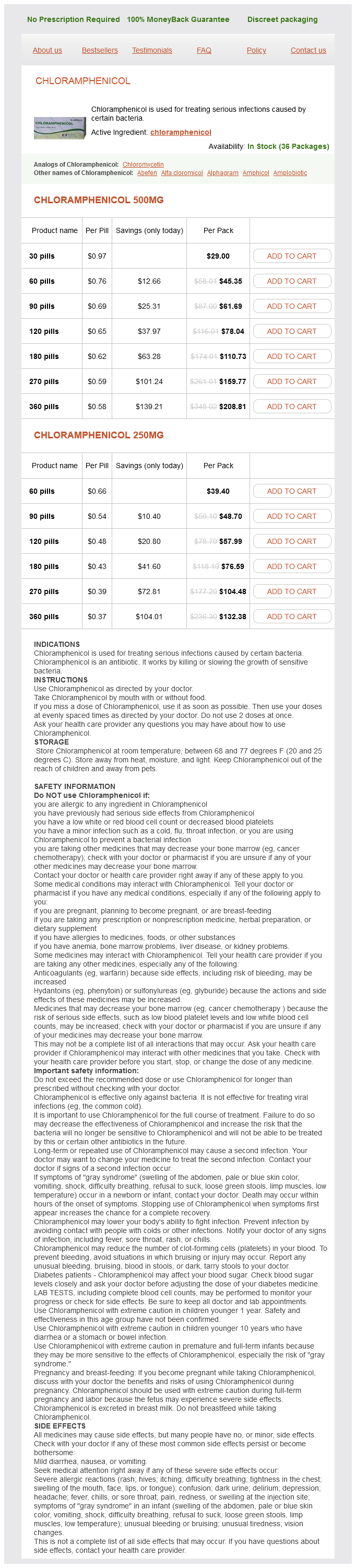
Chloramphenicol, also known as chloram, is an antibiotic medication broadly used for treating severe infections brought on by certain micro organism. This highly effective antibiotic is efficient towards a wide range of bacterial infections, making it a valuable software in the battle against infectious illnesses. In this text, we are going to discuss what chloramphenicol is, how it works, its uses, unwanted facet effects, and precautions.
Chloramphenicol is primarily used for treating severe infections attributable to bacteria corresponding to meningitis, sepsis, and typhoid fever. It can also be used to deal with infections of the attention, including bacterial conjunctivitis, and for treating certain kinds of pores and skin infections. In addition, it is efficient in treating bacterial respiratory infections, such as pneumonia and bronchitis.
In conclusion, chloramphenicol is a robust and effective antibiotic used for treating critical bacterial infections. When used appropriately and beneath the steering of a doctor, it could be a life-saving treatment. However, it is important to concentrate to the potential unwanted effects and take essential precautions whereas utilizing this medication. If you experience any extreme unwanted side effects, all the time consult your doctor immediately. With correct use and precautions, chloramphenicol could be a valuable weapon within the battle in opposition to bacterial infections.
It is essential to finish the complete course of the prescribed remedy, even when signs improve. Stopping the medicine early can result in the return of the an infection, and the bacteria may develop a resistance to the antibiotic.
In some cases, chloramphenicol could additionally be prescribed instead therapy for these who are allergic to other forms of antibiotics. However, it ought to only be used under the steerage of a physician as it is a highly effective treatment with potential unwanted facet effects.
Just before closing antibiotics to treat diverticulitis discount generic chloramphenicol canada, a small cut is made into the frenulum to bury the suture deep to the mucosa. Septospinal Suture If the septal base has the tendency to slip off the anterior nasal spine and premaxilla, the septal plate may be held in place by a septospinal suture. A slowly resorbable 3 suture is passed through the septal base, downward through the connective tissue fibers and the buccal mucosa on the left side of the anterior nasal spine and the frenulum of the upper lip, and then back through the mucosa and the connective tissue on the right side of the nasal spine. In reconstructing the septum, transseptal fixation sutures and septal splints may be of great help. The splints should not touch the mucosa of the floor or the roof of the nasal cavity. In special cases, they are left in place for a longer period of time, for example to prevent recurrence of synechiae. It is lifted upward and exorotated until the cartilaginous dorsum has reached the desired level. Fixation of the Septal Base in a Groove in the Premaxilla To stabilize the septum, a groove may be cut in the middle of the premaxilla with a 4-mm or 7-mm chisel. It leads to breathing obstruction on inspiration and sometimes also to cosmetic complaints because of the visible protrusion of the caudal septal end into the nostril. A vertical chondrotomy is made at the bend (fracture line) on the opposite side, and the septal base is disconnected from the anterior nasal spine and premaxilla. In cases with severe scarring, a unilateral or bilateral inferior tunnel may also be required (three-tunnel or four-tunnel approach). It only remains attached to the posterior part of the septum underneath the nasal dorsum. In spite of these resections, the anterior septum may tend to return to its dislocated position. Fixation of the caudal septal end by septocolumellar sutures and a septospinal suture. Such a graft is usually 20 to 25 mm long, somewhat longer than the height of the still intact more posterior part of the septum. If no septal cartilage is available, auricular cartilage is the second choice (see page 244 and page 350). In this case, the caudal end is resected and reconstructed by a cartilaginous graft (preferably autogeneic septal cartilage or auricular cartilage) that is positioned on the anterior nasal spine and premaxilla, and sutured to the intact posterior part of the septum. This graft corrects the retracted columella and the underprojected An incision (here, the term hemitransfixion would be correct! A columellar pocket is created in the membranous septum using small, sharp and blunt curved scissors. Care is taken not to extend the pocket superiorly to the upper (ventral) end of the nostril. The remaining caudal septum is carefully dissected up to its ventrocaudal corner, and then cranially up to the remaining septal cartilage. A rectangular piece of cartilage of sufficient size is now resected from the posterior part of the cartilaginous septum. Its length should be 20 to 25 mm, the distance between the anterior nasal spine and premaxilla and the domes. The graft should not be too thick to avoid broadening of the columella and narrowing of the valve area. When the convexity concerns the nasal valve area and inspiratory breathing is impaired, its correction is of utmost importance. A caudal convexity usually results from frontobasal trauma, and is often combined with asymmetry of the lobule. The cartilaginous septum is exposed and dislocated from its base and a small basal strip is resected. Sometimes such a cartilaginous convexity straightens out when the mucoperichondrium on both sides is elevated. Gridding or so-called cross-hatching of its concave side to break the elastic forces of the cartilage is often advocated. In combination with a batten graft (see below), some gridding of the concave side may be helpful. However, it weakens the cartilage and may thus cause sagging of the dorsum and retraction of the columella. A batten graft sutured to the concave side is more reliable and is therefore the method of choice. Batten Graft Steps Anterosuperior tunnels are made on both sides to completely expose the septal pathology. If the convexity does not straighten out by itself, a vertical chondrotomy is performed at the area of the maximal bending. A rectangular cartilaginous batten of the desired size is then harvested from the posterior part of the cartilaginous septum. It should bridge the distance between the anterior nasal spine and the nasal dorsum. The removed cartilage is extracorporally modified to a flat plate by conservative gridding, cross-hatching, or slight morcellation. Two or three guide sutures (3 resorbable vicryl) are fixed to the transplant: one at its future ventrocaudal corner; one just above its inferior caudal corner; and one through its dorsal margin. While the assistant is pulling the three guide sutures, the surgeon fixes the plate with two or three transseptal sutures. Positioning and fixing the autotransplant in its proper position is of utmost importance to avoid postoperative sagging of the cartilaginous dorsum. Although osteotomies may align the bony pyramid, the cartilaginous part will remain curved. Anterosuperior tunnels are made on both sides to completely expose the septal pathology.
The second applicator is positioned against the lateral nasal wall antibiotics used for sinus infections uk 250 mg chloramphenicol buy fast delivery, lateral to the tail of the middle turbinate. A third applicator may be applied on the nasal floor at the posterior end of the hard palate, where branches of Steps Infiltration (0. General anesthesia should be stabilized before additional local anesthesia and vasoconstriction can be applied. The level of the general anesthesia should remain the same until the very end of the operation. The patient is positioned with the head about 40 cm above the level of the feet (anti-Trendelenburg position). Additional local vasoconstriction and anesthesia are applied according to the rules that have been agreed upon with the anesthetist. The cocainedrenaline mixture may then be substituted by a local application of xylometazoline 0. Blocking the infraorbital and supraorbital nerves is not necessary in general anesthesia. In cases where time-consuming septal and lobular surgery has to be carried out, it is advisable to reinforce the paranasal infiltration before osteotomies are performed. Compression of soft tissues during the operation Additional Vasoconstriction and Local Anesthesia Endonasal Topical Anesthesia and Vasoconstriction Small gauzes soaked in a solution of cocaine 3% with epinephrine 1:20,000 are carefully applied endonasally in areas 5, 4, and 3. The gauzes are prepared on a separate table to avoid confusion with the topical anesthetic. Patient Position the patient is positioned with the level of the head about 40 cm above the feet. The head is also slightly rotated backward and positioned on a stabilizing cushion. Care is taken not to rotate the neck too much so as to avoid obstruction of venous backflow in the neck. In general, a systolic blood pressure of 100 to 110 mm Hg is usually sufficient to control bleeding when it is combined with the other measures discussed here. Local application of a cocaineadrenaline mixture to the nasal mucosa will prevent mucosal bleeding for about 1. Local infiltration of areas where incisions are planned with a lidocainedrenaline mixture greatly enhances a bloodless approach, while paranasal and infralobular infiltration is of great help in hump resection, osteotomies, and lobular surgery. Surgery in Anatomical Planes Dissection should be performed in anatomical planes as much as possible. Sometimes, cutting through tissues cannot be avoided, for example when making incisions, in hump removal, and when dissecting scar tissue. To avoid unnecessary tissue damage and bleeding, the septum is approached subperichondrially and subperiosteally, and the nasal dorsum is undermined at the level of the loose connective tissue layer just above the perichondrium and the periosteum. Lateral osteotomies are preferably performed subperiosteally, while medial osteotomies are made intraseptally. Compression of Tissues Compression of the tissues during surgery, especially in its final stages, is of great help in diminishing bleeding and preventing postoperative hematomas and edema. Temporary dressing of the nasal cavity after the first phase of septal surgery while working on the pyramid and lobule may also be of great help. Scher and Pirsig 1988, on the contrary, saw a distinctly higher rate of infectious complications in their study in septorhinoplasty patients in those who did not receive prophylactic antibiotics compared with those who did (5 versus 1 serious infection and 9 versus 3 moderate infections). Amoxicillin + clavulanic acid 500/125 3 dd 625 mg In combination with Flucloxacillin 4 dd 500 mg i. Weimert and Yoder 1980 concluded from their prospective randomized study that the incidence of infectious complications is not sufficient Corticosteroid schemes 1. When transplants (especially allogeneic or heterotopic) are used in reconstruction, the use of a broad spectrum antibiotic is indicated. This is not sufficient reason, however, to administer corticosteroids following nasal surgery except in special circumstances, such as patients with allergy or nasal polyposis. An incision is an opening made in the skin or mucosa that allows us to gain access to a certain area or anatomical structure. For instance, the caudal septal incision gives access to the septum, or the intercartilaginous incision serves as an entrance to the nasal dorsum. An approach is a surgical method used to arrive at a certain structure so that it may be modified. For instance, an anterosuperior septal tunnel is used as an approach to the cartilaginous septum, or undermining of the skin of the nasal dorsum is a means to approach a bony hump. A technique is a surgical procedure or method by which an anatomical structure is mobilized, repositioned, or modified. For instance, the let-down technique is used to lower a prominent pyramid, or the inversion technique is used to narrow the nasal tip. Generally speaking, a technique is a method by which tissues are repositioned, modified, or resected. External incisions are often said to become "almost invisible" when the tissues are handled delicately and sutured precisely. This may be true, but a completely invisible internal scar is always preferable to an almost invisible external one. For instance, the columellar inverted V incision is inevitable in the external approach.
Chloramphenicol Dosage and Price
Chloramphenicol 500mg
- 30 pills - $29.00
- 60 pills - $45.35
- 90 pills - $61.69
- 120 pills - $78.04
- 180 pills - $110.73
- 270 pills - $159.77
- 360 pills - $208.81
Chloramphenicol 250mg
- 60 pills - $39.40
- 90 pills - $48.70
- 120 pills - $57.99
- 180 pills - $76.59
- 270 pills - $104.48
- 360 pills - $132.38
Bilateral infracture requires paramedian antibiotics z pack and alcohol cheap chloramphenicol 500 mg online, lateral (or oblique), and transverse osteotomies on both sides. When a lateral osteotomy is done too high (too ventral), a visible and palpable "step" in the lateral bony wall may result. An external stent is applied with some pressure to keep the nasal bones in their new position. Endonasally, a limited amount of internal dressing is applied to avoid outward movement of the infractured bones by postoperative swelling. This can only be accomplished successfully when the pyramid is fully mobilized by performing bilateral paramedian, lateral, and transverse osteotomies, and mobilizing the septum. The long, shallow side is infractured, whereas the short, steep side is moved outward. The lateral osteotomy on the longer side is performed somewhat higher than on the short side. The distance between both osteotomies and the dorsum should be symmetrical so that the new lateral walls are equally high. To preserve the result, it is crucial to fix the bony walls and the septum in their new position. Rotation by Unilateral Wedge Resection Following mobilization of the bony pyramid and the septum, a wedge of bone is resected at the base of the long side of the bony pyramid. To stabilize the bony pyramid and the septum in their new position, internal dressings, external taping, and splinting are applied with special care. Bilateral outfracturing requires complete paramedian, lateral, and transverse osteotomies on both sides. Generally, however, the let-down procedure (see following text) is preferred in these cases. As previously noted, the lateral osteotomy should be done low to avoid a postoperative "step. Push-Down with Bilateral Infracture the bony pyramid is pushed down and bilaterally infractured. The technique 217 Pyramid Surgery requires resection of a basal horizontal and a posterior vertical strip from the septum in combination with bilateral paramedian, lateral, and transverse osteotomies. This method, introduced by Cottle (1954), may be used in patients with a prominent pyramid and a slight bony hump. Although elegant and conservative in comparison with other methods of hump removal, the technique has two disadvantages. In patients with a prominent pyramid, this is usually contraindicated, as their external nose and valve area are already narrow. In patients with a prominent, narrow nasal pyramid and hump, bilateral wedge resection followed by letdown of the pyramid is usually the best choice. The let-down technique requires resection of both a basal horizontal and a posterior vertical strip from the septum. It is therefore the technique of choice in patients with a narrow and prominent nasal pyramid with or without a bony and/or cartilaginous hump. The main ones are laceration of the endonasal mucoperiosteum, and production of bone dust. However, it is difficult to bring the nasal bones upward and keep them in a more ventral position. External fixation is the only effective means to preserve a push-up of the pyramid. There is no consensus on which type of chisel is the best; choice is based largely on personal preference. Chisel the chisel is nowadays the preferred instrument for performing osteotomies. Accordingly, the two sides meet a different amount of resistance while proceeding. When the bevel is facing up, the bone cut will be convex; when the bevel is facing down, it will be convex. Nowadays, Saw In the early era of nasal surgery, lateral osteotomies were performed with a saw. In the 1950s, Cottle introduced a greatly improved design with a hand grip and a changeable band saw (see Chapter 10). For beginners, the guided chisel, as advocated by Masing, may be a good alternative for performing lateral osteotomies. We have a preference for these chisels, as they are universal and can be used intraseptally as well as for osteotomies and hump resection. The 4-mm and the 7-mm chisel are used for resecting crests and spurs, as well as for making cuts into the vomer and the perpendicular plate. The curved 6-mm chisel is used to make a vertical cut into the perpendicular plate to resect a bony plate for transplantation. The 7-mm straight chisel is the instrument of choice for paramedian and lateral osteotomies, as well as for wedge resections. By choosing between "bevel up" and "bevel down," the bone cut can be made along the required course. The curved 6-mm chisel is used for transverse osteotomies, while the 9-mm and 12-mm chisels are taken for hump resection. A disadvantage of Cottle chisels is that beginners may find them somewhat difficult to guide.

ADDII BIOTECH is proud to announce that the company's manufacturing facility located at Baddii, Himachal Pradesh has recently been approved by Food & Drug Authority of Ghana.
Our Manufacturing capability includes a wide range of therapeutic products covering almost every segment.
ADDII BIOTECH given our strong emphasis on product quality and services.