
Cialis with Dapoxetine
General Information about Cialis with Dapoxetine
By combining these two medications, Cialis with Dapoxetine effectively addresses both ED and PE, permitting males to achieve and keep a longer-lasting erection whereas also delaying ejaculation. This combination medication is available in numerous dosages, giving men the flexibleness to determine on the best strength that works for them.
One of the significant advantages of Cialis with Dapoxetine is its convenience. Instead of taking two separate medicines, men can take just one pill of Cialis with Dapoxetine roughly one to three hours earlier than sexual activity. This comfort not only saves time but in addition makes it simpler for men to adhere to their treatment plan.
However, as with all medicine, there may be side effects. The most common unwanted effects of Cialis with Dapoxetine embrace headache, dizziness, nausea, and flushing. These side effects are usually mild and well-tolerated. It is always essential to seek the assistance of with a healthcare skilled earlier than starting any new medication.
Erectile dysfunction (ED) and premature ejaculation (PE) are two frequent sexual well being points that may significantly affect a man's confidence and relationship with their companion. While they're two distinct issues, they often occur collectively, making it difficult to search out an efficient remedy. However, with the introduction of Cialis with Dapoxetine, also identified as Super Tadarise, males now have an efficient and convenient solution to combat each ED and PE.
Cialis, with its energetic ingredient Tadalafil, is a popular treatment used to treat ED. It works by enjoyable the muscular tissues in the walls of the blood vessels, permitting increased blood move to the penis, resulting in a firmer and longer-lasting erection. This effect lasts for up to 36 hours, making it a well-liked choice amongst males.
In conclusion, Cialis with Dapoxetine is a game-changer within the remedy of ED and PE. Its mixture of Tadalafil and Dapoxetine provides a complete resolution for males fighting both conditions. It is handy, effective, and well-tolerated, making it a preferred alternative amongst males looking to enhance their sexual well being. However, it is crucial to observe dosage directions and consult a healthcare professional to ensure protected and efficient use of this treatment.
ED is a condition the place a person is unable to achieve or maintain an erection essential for sexual intercourse. It can be brought on by various factors corresponding to psychological issues, life-style decisions, or underlying medical conditions. On the other hand, PE is characterized by a man ejaculating too early, often earlier than or inside a minute of penetration. It can additionally be brought on by psychological elements, physical trauma, or hormonal imbalances.
Cialis with Dapoxetine is a combination medication that incorporates two active ingredients - Tadalafil and Dapoxetine. Tadalafil, also referred to as Cialis, is a drugs used to treat ED, whereas Dapoxetine is a drugs used to treat PE. When each these components are combined, they work synergistically to supply a potent remedy for men battling both ED and PE.
Dapoxetine, the opposite lively ingredient in Cialis with Dapoxetine, is specifically used to treat PE. It belongs to a category of medications often identified as selective serotonin reuptake inhibitors (SSRIs). It works by increasing the degrees of serotonin within the brain, which helps to delay ejaculation and enhance management over ejaculation.
A multiinstitutional trial published in 2002 reported an overall upgrade rate of 8% at surgical excision (34) erectile dysfunction at age 18 cheap cialis with dapoxetine 20/60 mg on line. There was no upgrade if at least 12 specimens were obtained at stereotactic biopsy using an11-gauge vacuumassisted device, if there was no associated atypia, and when the mammographic findings were concordant. Microscopic radial scars also have a low upgrade to malignancy and may not require excision (35). Although the data are limited, some institutions reserve surgical excision for radial scars that are mammographically evident or for radial scars associated with atypia. Fine-needle aspiration biopsy of nonpalpable breast lesions in a multicenter clinical trial: results from the radiologic diagnostic oncology group V. Probably benign breast lesions: when should follow-up be recommended and what is the optimal follow-up protocol Solid breast nodules: use of sonography to distinguish between benign and malignant lesions. Probably benign lesions at breast magnetic resonance imaging: preliminary experience in high-risk women. Automated large-core needle biopsy of surgically removed breast lesions: comparison of samples obtained with 14-, 16-, and 18-gauge needles. Benign Papillomas Papillary lesions are diverse, ranging from benign papillomas to papillary lesions with atypia to invasive papillary carcinomas. However, there is continuing controversy as to whether surgical excision is required for a benign papilloma on core biopsy. Surgical excision has traditionally been recommended due to the limited sample obtained and concern that percutaneous biopsy might sample a nonrepresentative portion of the lesion. Similar to other high risk lesions, the reported upgrade rate of papillomas ranges widely in the literature between 0% and 25%, largely due to the small number of patients included in each study, their retrospective design, and differences in assessing imaging-pathologic concordance (31,36). Most of these studies involved papillomas diagnosed at either ultrasound or stereotactic biopsy. Communication with the pathologist is important to distinguish microscopic papillomas that are entirely contained within the core specimens as these may not require excision (35). Percutaneous core biopsy is a safe, less invasive, and less costly alternative to surgical biopsy with comparable accuracy. However, understanding potential pitfalls that may occur with image guided biopsy is important in order to maintain its effectiveness. Accurate targeting is critical with any image guided biopsy in order to minimize false negative rates. Management of high risk lesions continues to be debated, and additional studies need to be performed to standardize management of these lesions. Utility of 6-month follow-up imaging after a concordant benign breast biopsy result. Controversies on the management of highrisk lesions at core biopsy from a radiology/pathology perspective. Atypical lobular hyperplasia and classic lobular carcinoma in situ in core biopsy specimens: routine excision is not necessary. Percutaneous core needle biopsy of radial scars of the breast: when is excision necessary Risk of malignancy when microscopic radial scars and microscopic papillomas are found at percutaneous biopsy. These advances are based on the discovery and characterization of a number of high-risk, relatively uncommon genes responsible for the clustering of breast cancer in certain families. More recently, a large number of common variants having a modest effect on individual risk have been defined by the use of genome-wide association studies. As clinical utility is currently largely restricted to high-risk genes, this chapter will focus largely on this category but in the future it seems possible that low-risk common variants will also be utilized to inform risk and management of breast cancer. Although genetics are clearly important, there is a tendency to assume that familial clustering of disease invariably results from inherited predisposition. However, other explanations for familial clustering of breast cancer should be considered including (a) geographically limited environmental exposure to carcinogens, which might affect an extended family living in close proximity; (b) culturally motivated behavior that alters risk factor profile, such as age at first live birth; and (c) socioeconomic influences that, for example, might result in differing dietary exposures. In addition, multiple, complex inherited genetic factors likely influence the extent to which a risk factor for breast cancer plays a role in any one individual; such modifying effects are likely to be shared among genetically related members of an extended family. Although many of these studies have methodological flaws, they consistently demonstrated a twofold to threefold increase in breast cancer risk in mothers and sisters of patients with breast cancer. The first large population-based study to estimate breast cancer risk associated with a family history was conducted in Sweden and involved 2,660 women (3). Within this study cohort, women with an affected relative had an increased breast cancer risk of 1. Anderson (4) suggested that a small subset of families with a very high risk of developing breast cancer due to a single genetic defect might be obscured in studies in which most breast cancer cases were multifactorial in origin. In 1984, Williams and Anderson (5) provided the first evidence for an autosomal dominant breast cancer susceptibility gene with age-related penetrance finding supported by Newman et al. With a pattern of autosomal dominant inheritance, an individual can have one of three possible genotypes: carrier of two nonmutant alleles (homozygous normal), or carrier of one (heterozygous) or two (homozygous) mutant alleles. The actual risk of developing breast cancer in a mutation carrier is based on the penetrance of the gene. Penetrance is the likelihood that the effect (phenotype) of a mutation (genotype) will become clinically apparent. Individuals carrying two copies of an autosomal dominant diseaserelated gene are rare, partly because of the relative rarity of heterozygotes and partly because of the potential for a lethal defect in a homozygous affected fetus. Anecdotal observations suggest that these women develop more frequent and earlier cancers than single mutation carriers, but the number of such individuals identified is too small for definitive studies. There is a 50% chance that an individual offspring will inherit a mutant copy of any given gene from a heterozygous parent. Therefore, on average, 50% of the related individuals in a family carry the mutant gene being transmitted. If the penetrance of the gene is high, the pedigree pattern for an autosomal dominant disease is quite striking, with vertical inheritance and half the children of an affected parent also being affected, whereas none of the offspring of a homozygous normal parent are affected.
When both tests were negative erectile dysfunction caused by vascular disease discount cialis with dapoxetine 20/60 mg otc, the specificity of the ultrasound exam improved from 25% to 34% (p <. They concluded that an anatomically low suspicion mass with a negative elastogram and Doppler interrogation could be called probably benign and undergo 6-month follow-up rather than biopsy (21). It works by measuring the propagation of the speed of the sound waves which is directly related to tissue stiffness. The device is placed over the breast and static images are obtained in a standard fashion which also allows 3-D reconstruction. This may improve efficiency, but patients will need to be called back if there are findings requiring additional evaluation. Pathology revealed grade 1 infiltrating ductal carcinoma with ductal carcinoma in situ. However, specificity remains suboptimal due to demonstration of vascularity within benign tumors. Gas microbubbles within lipid microspheres have been injected in an attempt to improve sensitivity and specificity of ultrasound. The microbubbles oscillate and emit signals that can be detected by the ultrasound probe. This type of contrast is different from iodinated contrast in that the microbubbles do not diffuse from the blood vessels into surrounding tissues. Newer ultrasound imaging techniques have been developed to better image these microbubbles. Early studies have shown improved sensitivity of up to 100% using this technique, but with specificities of 5. Limitation of specificity is likely due to increase in the detection of small, nonmalignant vessels. Possible applications include the follow-up of patients after neoadjuvant chemotherapy or as a method to deliver drugs directly to a tumor site. While potentially very exciting, the use of microbubbles is currently limited due to the technical difficulty involved in performing this procedure. Cost is high due in part to contrast material, length of time for examination, and interpretation. Interpreting the first postcontrast subtraction views took 58 seconds to read with a sensitivity of 98. When interpreted with no history or prior examinations, one reader detected 98% of the cancers on the first postcontrast images and first postcontrast subtraction images while the second reader detected 95% and 93%. The time to perform these limited sequences is approximately 15 minutes, reduced from the full protocol which takes 30 to 40 minutes. This is a promising area of exploration, although a great deal more work needs to be done. With regard to sensitivity, it is the examination against which all other breast imaging exams are compared. Images are obtained prior to and at several time points after infusion of a gadolinium chelate contrast material (except when evaluating implants). Lesions with irregular, microlobulated, or spiculated margins are more likely to be malignant. Segmental or ductal distribution of nonmass enhancement also increases the likelihood of malignancy. Rapid initial enhancement followed by washout of contrast is more highly suggestive of carcinoma compared to a plateau or continual increase in enhancement. It is a pulse sequence that essentially measures the random motion of water molecules within a lesion. Motion is affected by cellularity and extracellular characteristics such as viscosity, membrane permeability, and blood flow. They demonstrated an improvement in the area under the receiver operating curve from 0. This function is based on the fact that choline is a precursor of phosphatidylcholine which composes cell membranes and increases with tumor growth. It has been well established that choline peaks can be detected in most breast cancers and generally not in normal breast tissue. There is, however, some overlap in choline values between benign and malignant tumors. In their evaluation of 208 breast lesions (169 malignant and 39 benign), sensitivity decreased from 84. They also found that both results correlated significantly with nuclear grade, estrogen receptor negativity and triplenegative lesions (32). Lipid signals from the surrounding adipose tissue may contaminate this measurement and precise placement requires the breast imager to be available to determine the area to be measured at the time of scanning which can hamper use in a busy clinical setting. Measurements in lesions under a centimeter are likely to be inaccurate limiting use in both the diagnostic setting as well as the evaluation of residual disease after treatment. They have shown that there is less likely to be contamination from surrounding tissues using this technique with the different metabolites which would therefore not require such precise identification of the area to be measured. Sensitivity was excellent (greater than 90%) for large lesions but abysmal for small lesions. There does not appear to be a significant clinical difference between the two technologies.
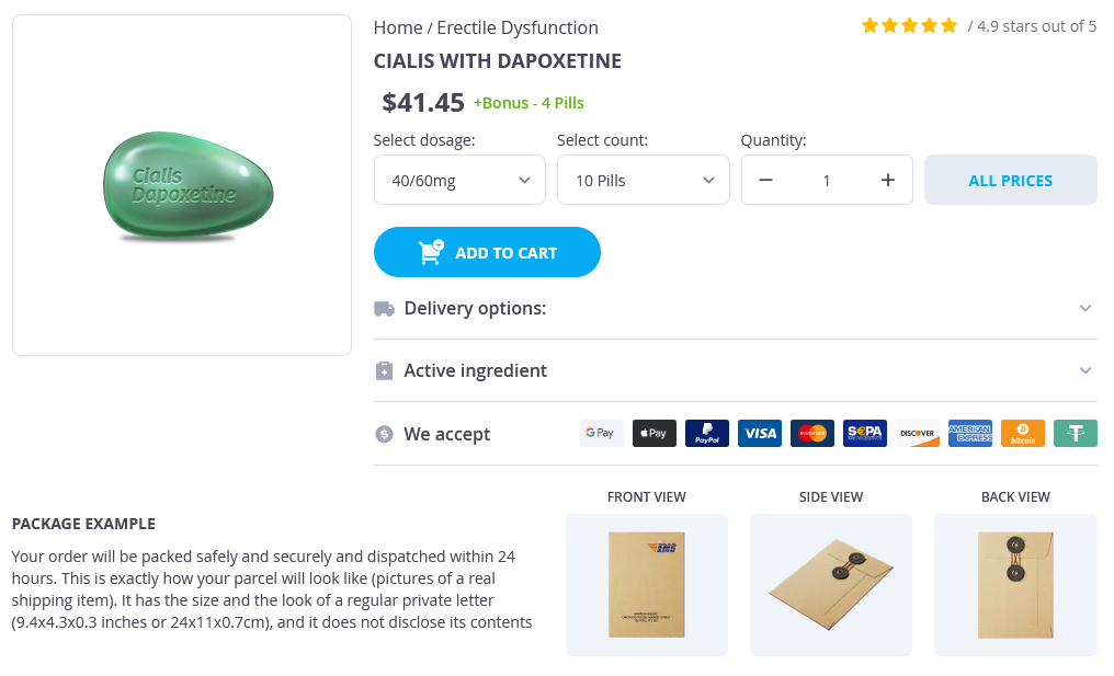
Cialis with Dapoxetine Dosage and Price
Cialis with Dapoxetine 40/60mg
- 10 pills - $46.05
- 30 pills - $124.06
- 90 pills - $296.09
- 120 pills - $372.02
Cialis with Dapoxetine 20/60mg
- 10 pills - $42.06
- 30 pills - $108.09
- 90 pills - $276.07
- 120 pills - $342.05
- 180 pills - $455.06
Classification of injuries is discussed how erectile dysfunction pills work buy 20/60 mg cialis with dapoxetine free shipping, as this provides the framework for planning repairs; it is also important for documentation of cases and communication among health care professionals. With classification understood, it is then important to know how to define the injuries so that they can fit into the classification system, so diagnostic testing and evaluation are discussed. In this regard, it is critically important to remember to assess patients for associated injuries, particularly the cervical spine, as the presence of an unrecognized cervical spine Classification of Nasal and Paranasal Sinus Trauma To understand and discuss sinus trauma, we need to have some definitions and categories that will help clarify the injuries involved and allow communication among surgeons. The Le Fort classification system is based on work done by Renee Le Fort that was originally published in 1901. Note that, although most facial fractures are often more complicated than those represented in the Le Fort classification system, the categories that he defined still provide an excellent framework for the basic patterns of fractures that occur. More importantly, the Le Fort definitions are commonly understood by most specialists/surgeons who treat maxillofacial fractures. This system therefore provides an excellent basis for communication among providers who care for these patients. The presence of teeth makes fracture repair more difficult, as it decreases the tolerance in the system for imperfections in fracture reduction. Patients may be quite sensitive to minor discrepancies in tooth position, and, of course, significant discrepancies translate into malocclusions that often lead to functional impairment and temporomandibular joint problems. Other potential pitfalls result from the presence of multiple visceral structures that are placed at risk from both the injuries and the repairs, including the eyes, the brain, salivary glands and ducts, and multiple sensory and motor nerve branches, as well as skin and muscle problems that may cause cosmetic and functional problems. When combined with a Le Fort fracture, it makes reestablishing the occlusion more difficult and therefore adds to the complexity of the repair. It also allows for the occurrence of a unilateral Le Fort fracture, seen most commonly at the Le Fort I level. Dental Arches (Alveoli) Fractures through the dental arches may occur independently or together with other fractures. When they occur in isolation, they are usually managed by either dentists or oral/maxillary surgeons. When they occur together with Le Fort fractures, their presence makes the repair more difficult, as the dental occlusion may move independently of the maxillary bones, making it more difficult to identify the proper relationship of the bone fragments. It crosses the nasal septum and is completed posteriorly by fracturing through the pterygoid plates (an important component of all true Le Fort fractures). Obviously, the fracture involves the maxillary sinuses, though it generally avoids the ostia of the sinuses and therefore should not be expected to alter sinus function. Like the Le Fort I, the fracture crosses the maxillary sinuses, nasal septum, and pterygoid plates. However, instead of a reasonably horizontal orientation, the fracture extends from the lateral maxillary sinus in a more superior direction through the inferior orbital rim into the orbit, involving the orbital floor. It then extends across the medial orbital wall and involves the nasal bones, classically separating them from the frontal bones at the nasal root. Splitting of the palate usually occurs in a Nose Nasal Bones Various classifications have been proposed for nasal bone fractures, although few surgeons use them. Most discussions of nasal fractures are therefore descriptive, rather than based on classification. In analyzing nasal injuries, once issues of bleeding and infection have been addressed, the key concerns are appearance and function. The presence of a preexisting deformity, cosmetic and functional, will influence the decision of whether or not to consider an attempt at acute repair and by what means. Although the sense of smell can be adversely affected by nasal fractures, most functional complaints relate to nasal airway obstruction. Classification of Nasal and Paranasal Sinus Trauma 547 Nasal Septum the nasal septum is frequently fractured and/or dislocated as a result of nasal trauma. It is difficult to estimate the frequency of this injury, as most people probably do not even seek treatment for minor nasal traumas. However, if a patient presents with nasal trauma, it is most important to examine the nasal septum to rule out the presence of a septal hematoma, as failure to identify and manage this problem can lead to long-term sequelae. A septal hematoma develops in the plane between the perichondrium and the septal cartilage. Because the cartilage derives its blood supply from the perichondrium, failure to identify and manage the hematoma can lead to cartilage necrosis and/or abscess formation, which can lead to both airway problems and cosmetic (saddle nose) deformity or worse. Classification of this fracture is typically based on the status of the bone fragment that bears the medial canthal ligament, rather than the status of the other fractured bones. The solid malar eminence usually remains fairly intact, whereas the thinner connections that stabilize the eminence in space usually break. These include the thin zygomatic arch, which articulates with the temporal bone and the thicker lateral orbital rim. Fracture of these bony attachments allows the eminence to rotate, usually medially and inferiorly, although it can rotate laterally as well, and sometimes directly inward. The third and fourth attachments are continuous, explaining the terms tripod and tetrapod, and the solid attachments are the inferior orbital rim and the lateral buttress between the zygoma and the maxilla. The fracture traverses the generally thin anterior maxillary bone and the thin orbital floor and inferolateral wall. Orbits Orbital Floor (Roof of the Maxillary Sinus) the floor of the orbit is the roof of the maxillary sinus. Fractures are categorized as blowout fractures, which are fractures of the orbital floor that occur in association with an intact orbital rim, and other orbital floor fractures, which occur in association with rim fractures, along with maxillary and zygomaticomaxillary fractures. Some experts argue that certain traumas to the rim will cause it to buckle without fracturing, leaving a blowout fracture of the orbital floor. Others believe that direct trauma to the globe results in transmission of force to the orbital floor, causing it to fracture. The classical pattern crosses the orbit above the infraorbital rim, fracturing across both the medial and lateral orbital rims and walls, while also crossing the upper nasal bones or nasal root. The craniofacial separation is completed by disarticulating the zygoma from the temporal bone via fractures through the zygomatic arches.

ADDII BIOTECH is proud to announce that the company's manufacturing facility located at Baddii, Himachal Pradesh has recently been approved by Food & Drug Authority of Ghana.
Our Manufacturing capability includes a wide range of therapeutic products covering almost every segment.
ADDII BIOTECH given our strong emphasis on product quality and services.