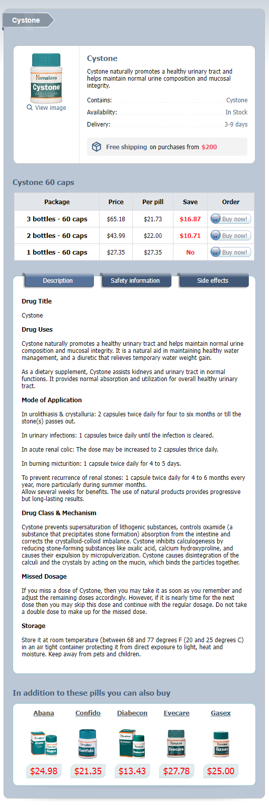
Cystone
General Information about Cystone
Urinary tract well being is an important aspect of overall well-being. It performs a vital position in eliminating waste products and toxins from the physique and maintaining a balanced pH stage. However, various components similar to dehydration, poor diet, insufficient hygiene, and bacterial infections can disrupt the normal functioning of the urinary tract. This can result in a bunch of uncomfortable and often painful conditions, together with urinary tract infection (UTI) and kidney stones. Fortunately, nature has offered us with a strong herbal remedy that may help keep a wholesome urinary tract � Cystone.
Cystone has been extensively researched, and its effectiveness has been supported by clinical studies. One research printed in the Journal of Clinical and Diagnostic Research showed that Cystone has important antibacterial and anti inflammatory exercise, making it an efficient pure treatment for UTIs. Another examine published within the International Journal of Advanced Ayurveda, Yoga, Unani, Siddha, and Homeopathy demonstrated the antiurolithiatic (kidney stone preventing) effects of Cystone in lowering stone formation.
In conclusion, Cystone is a natural, protected, and effective approach to promote a healthy urinary tract. Its potent elements work synergistically to prevent and deal with UTIs, promote regular urine composition, and preserve mucosal integrity. Moreover, it is free from any antagonistic unwanted effects and may be safely used as a long-term answer to hold up urinary tract concord. So, for anyone seeking to naturally assist their urinary tract health, Cystone is certainly a supplement to think about.
Cystone is a herbal supplement that has been used for tons of of years in Ayurvedic medication to advertise urinary tract health. It is a proprietary mix of highly effective components, including Shilapushpa (Didymocarpus pedicellata), Pasanabheda (Saxifraga ligulata), and Manjishtha (Rubia cordifolia), among others. These natural components work collectively to help the normal functioning of the urinary tract by sustaining proper urine composition and mucosal integrity.
In addition to its benefits for the urinary tract, Cystone also has a diuretic effect. It helps improve the volume and frequency of urination, which is crucial for flushing out toxins and stopping the buildup of harmful bacteria. The elevated urinary circulate also helps forestall the formation of crystals and stones by diluting the urine and rising its pH stage.
One of the necessary thing benefits of Cystone is its capability to stop and treat urinary tract infections. UTIs are brought on by the overgrowth of bacteria in the urinary tract, leading to irritation and infection. Cystone incorporates anti-microbial properties that help combat the dangerous bacteria and stop their adherence to the walls of the urinary tract. This, in flip, helps prevent the unfold of an infection and promotes the healing of infected tissues.
Another essential side of urinary tract health is maintaining the integrity of the mucosal lining of the urinary tract. The mucosal lining helps defend the urinary tract from the harmful results of bacteria and toxins. When this barrier is compromised, it could lead to inflammation and an infection. Cystone incorporates anti-inflammatory and antispasmodic properties that help soothe and protect the mucosal lining, promoting its natural healing course of.
Furthermore, Cystone additionally supports normal urine composition. It helps stop the formation of crystals and stones within the urinary tract. This is very helpful for people who discover themselves susceptible to kidney stones. The potent ingredients in Cystone inhibit the growth and accumulation of minerals in the urinary tract, thus decreasing the chance of stone formation. Additionally, it also helps dissolve small stones, making them easier to move naturally.
The impact of the condition goes beyond the physical disability and psychological effects for the child to affect the entire family in terms of financial and social implications medications requiring central line cystone 60caps low price. Equally important in prevention is prenatal screening during pregnancy as well as the role of genetic counseling for future pregnancies. There appears to be a genetic predisposition and mutations in the folate responsive or folate dependent pathways may play a role. Some cells differentiate and migrate to form the neural crest on the lateral surface of the neural tube. The neural folds bend inwards due to changes in the shapes of neural cells to bring the tips of the folds into apposition. Then there is fusion of the tips of the folds leading to formation of the neural tube. The closure of the neural tube initially occurs in the center followed by rapid closure both rostrally and caudally. The vertebral column develops around the central notochord which develops from the notochordal plate. Those who exceed a threshold liability due to genetic and environmental factors, develop the trait while those below the threshold do not. But the recurrence risk still remains low at 3%, much lower than traits caused by a single fully penetrant mutation. Therefore, these defects arise from an interplay of a number of genes along with gene-environmental interactions. Evaluation of multiple genes may be required in an individual to determine risk as well as appropriate preventive therapy. The incidence varies in different parts of the world and different regions within countries. In United States of America, incidence is 1/1,000 births whereas it is as high as 12/1,000 births in some parts of Ireland and Wales. Craniorachischisis and iniencephaly are much more frequently seen in Northern China. The risk of recurrence in the next pregnancy is 4% which increases to 10% and 25% with subsequent pregnancies. Some other risk factors studied but not established are gestational diabetes, chlorination of drinking water, electromagnetic fields, pesticides and hazardous wastes. The success of folic acid supplementation is because folate metabolism contributes to many biological functions. MaternalAutoantibodies Neural tube defects can occur in babies of women who had maternal antibodies against folate receptors. In future, this may lead to clinical testing in first pregnancies to adjust the dose of folic acid supplementation in subsequent pregnancies. The clinical presentation depends on the stage of the embryological defect in the process of development of neural tube Table 1). Encephalocele is formed when there is herniation of brain and meninges through defect in calvaria. Craniorachischisis is defined as anencephaly associated with continuous bony defect of spine with exposure of neural tissue. The exact mechanism for increased risk is not known but this happens only when there is a genetic predisposition to the teratogenic effects of valproate. The anomaly consists of a midline defect of the vertebral bodies without involvement of spinal cord or meninges seen usually in the lower lumbar region at L5 S1 levels. The patients are asymptomatic with no neurological signs and the anomaly is detected accidentally on radiographic studies. The roentgenogram of the spine reveals a defect in closure of the posterior vertebral arches and laminae. It has been suggested that some host factors which are derived from mesoderm are essential to induce neurulation. The neural tube is patterned along three axes namely anteroposterior, dorsal-ventral and right-left axis as a response to the expression of intrinsic patterning genes and embryonic signaling pathways. Homeotic Hox and family transcription factors affect segmental identities and hindbrain features. These substances when found in amniotic fluid serve as biochemical markers of a neural tube defect. Dermal Sinus A small opening on the skin leads into a narrow passage which may be a dead end or pass through the dura and act as midline cutaneous makers over the location of a meningocele or encephalocele and are found in the lumbosacral or occipital region. Recurrent meningitis of unknown origin should alert one to look for a small dermal sinus. Associated conditions such as diastematomyelia and tethered cord should be looked for and require surgical intervention. In most cases, the spinal cord is normal but sometimes there can be tethered cord, syringomyelia or diastematomyelia. Transillumination is positive as there is no nervous tissue present in the lesion. The spectrum of clinical manifestations varies from severe sensory loss and trophic ulcers to only mild sphincter disturbances. Although intelligence is normal in the majority, cognitive and language difficulties may be present. The nature and severity of the neurological impairment depends on its location and size. Additionally, overflow incontinence, loss of sacral tone, loss of sacral and rectal sensations and loss of det usor muscle activity occurs. The clinical features are progressive hydrocephalus, secondary brainstem dysfunction, feeding and respiratory complaints including apnea. In 50Â85% of patients, aqueductal stenosis or atresia may also contribute or lead to the hydrocephalus.
Splinter hemorrhages are nonblanching medications 4h2 60caps cystone order otc, linear, reddish-brown lesions found under the nail bed. Septic embolizations are not uncommon and result in small or major arterial occlusions with infective infarctions in extracardiac organs. Community acquired Staphylococcus aureus or enterococci, in the absence of a primary focus or B. New valvular regurgitation (worsening or changing of pre-existing murmur not sufficient) Minor criteria 1. Vascular phenomena: Major arterial emboli, septic pulmonary infarcts, mycotic aneurysm, intracranial hemorrhage, conjunctival hemorrhages, and Janeway lesions 4. Routinely, three blood cultures (2Â3 mL of blood) under strict aseptic precautions are recommended in children. In 90% of cases of endocarditis, the causative agent is recovered from the initial two blood cultures. If there is no growth by 2nd day, two more blood cultures may be drawn in case antibiotics have not been started. Unless otherwise suspected, blood cultures are usually performed for aerobic organisms. Delayed cultures especially on enriched media are necessary to detect fastidious bacteria or fungi. If the patient is not acutely ill, antibiotic therapy can be withheld for 24Â48 hours while the blood cultures are obtained. The probability of pathogenicity is increased if the organism is observed in multiple blood cultures obtained by independent venipunctures. The limitations of echocardiography are: (1) False negative results with small vegetations or in embolized vegetations; (2) not all echogenic masses are necessarily vegetations; and (3) inability to differentiate active vegetation from an old sterile vegetation. Once treatment is completed, a repeat evaluation may be necessary to establish a new baseline of valvar and myocardial functions for the patient. Other abnormal echocardiographic findings not fulfilling the definitions are considered as minor criteria. The diagnostic yield of echocardiography is influenced by the image quality, size of vegetation, (vegetation < 2Â3 mm may not be well seen), location of vegetation. Urinalysis shows hematuria, proteinuria, and red cell casts suggestive of glomerulonephritis. Bactericidal antibiotics should be started empirically after blood cultures are collected, if the patient is acutely ill. However, if the child is stable, it would be advisable to wait for 48 hours till antibiotic sensitivity testing is carefully reviewed. Use of recombinant tissue plasminogen activator to lyse intracardiac vegetations in severely ill infants has been mentioned in literature. Digitalis, salt restriction, diuretic therapy and other standard protocol should be instituted in supportive care of heart failure. Duration of therapy is prolonged and generally for 6Â8 weeks, followed by oral antifungal agents like fluconazole. However, the superiority of two drug combination over amphotericin B alone has not been proven by randomized trial. Medical therapy alone is often not successful and surgical intervention is often necessary. Empiric Antibiotic Treatment It is generally directed to the most common pathogens, i. The chosen antibiotics must be given intravenously to attain persistently high bactericidal concentrations. Duration of therapy in patients with native valve endocarditis ranges from 2 weeks to 6 weeks, depending on the pathogen and site of valvar infection. Infection of prosthetic valves and tissue, infection with highly virulent or more resistant pathogens may require longer treatment usually 6Â8 weeks. Prolonged therapy is often necessary to eradicate organisms that are growing in relatively inaccessible avascular vegetation. The duration of penicillin-resistant therapy for streptococcal endocarditis on a prosthetic valve is 6 weeks. Susceptible enterococcal infection on native valves Penicillin or ampicillin, combined with gentamicin, for 4Â6 weeks. Susceptible enterococcal infection on prosthetic material should be treated for at least 6 weeks. Linezolid or daptomycin are options for patients with intolerance to vancomycin or resistant organisms. Substitution of linezolid for vancomycin should also be considered in patients Cardiovascular Disorders Surgical Intervention Surgical intervention should be contemplated early if the likely result is more favorable than with medical management, based upon an echo-guided analysis of the relative risks and benefits. The goals of surgery are to eradicate the focus of infection, to repair cardiac defects and to prevent development of complications. Postoperatively, a full course of antimicrobial therapy starting from the time of surgery is warranted and at times antibiotics changed according to culture sensitivity of organisms grown on operative valve/tissue cultures. Cardiac complications include cardiac failure, perivalvar abscess, pericarditis, and intracardiac fistula. Congestive heart failure can occur secondary to ruptured leaflets or chordae and is the most common cause of death. Development of a new onset heart block may suggest perivalvar extension of infection. Septic emboli can occlude or damage any vessel in the systemic or pulmonary arterial circulation. Following highest risk procedures may result in transient bacteremia: · All dental procedures that involve manipulation of either gingival tissue or the periapical region of teeth or perforation of the oral mucosa would warrant antibiotic prophylaxis.
Cystone Dosage and Price
Cystone 60 caps
- 3 bottles - $65.18
- 2 bottles - $43.99
- 1 bottles - $27.35
Depending upon the extent of the fold symptoms 0f pregnancy buy cystone 60 caps otc, it is referred to as epicanthus supraciliaris (eyebrow to lacrimal sac), epicanthus palpebralis (upper eyelid to inferior orbital margin), epicanthus tarsalis (upper eyelid tarsal region to medial canthus), and epicanthus inversus (from lower eyelid to medial canthus). This may occur in isolation or be associated with other abnormalities like ptosis, ankyloblepharon, telecanthus (Blepharophimosis syndrome). However, when treatment is indicated for cosmetic reasons, surgical correction using a V-Y procedure or Mustardes four flap technique is employed. Anophthalmia and microphthalmia may occur in isolation or in association with systemic disease. Telecanthus At birth, normal inter medial canthal distance is 20 ± 2 mm and normal interpupillary distance is 39 ± 3 mm. Telecanthus refers to an increased intermedial canthal distance, with normal interpupillary distance. It may occur in isolation or as an association with epicanthus and blepharophimosis. Genetics of Lacrimal System Congenital alacrima, a rare autosomal dominant condition is caused by mutations in the gene encoding (5p12). Blepharophimosis Narrowing of the palpebral aperture is referred to as blepharophimosis. Depending on the amount of eyelid deformity, patients undergo a staged repair of the eyelid defects. Ankyloblepharon Partial or complete fusion of the eyelid margins, leading to shortening of the palpebral aperture is referred to as ankyloblepharon. When the fusion of the two lids is by a single or multiple tags, it is referred to as ankyloblepharon filiformadnatum. Ectodermal defects, cleft lip/palate, hydrocephalus or meningomyelocele may be associated. Euryblepharon It is a congenital generalized enlargement of palpebral aperture, lateral end more than medial. The wider lateral canthus is associated with inferior displacement of lower eyelid (mimicking ectropion). Occurrence of euryblepharon may be isolated or inherited as an autosomal dominant trait. Treatment involves removal of the extra row of eyelashes either surgically or by using electrocautery/cryotherapy. Entropion Congenital entropion is the inturning of lower eyelid margin, resulting in epiphora due to corneal irritation. Entropion associated with epicanthus or epiblepharon, results from a hypertrophic orbicularis oculi muscle and is often familial. Surgical correction by spindle excision of skin muscle complex is treatment of choice. The anterior lamellar shortening is part of the generalized tightening of the skin as in Collodion babies. Lubricants and emollients are used on the skin to result in softening of the stretched tissues. Sometimes grafting (to increase the anterior lamella) may be required in persistent cases. Ptosis resulting from congenital third cranial nerve palsy or Horners syndrome is not conventionally referred to as congenital ptosis. Compensatory chin elevation occurs in moderate/severe bilateral cases, frontalis muscle overaction compensates unilateral cases. Ptosis is graded as mild, moderate or severe depending on the amount of levator action and palpebral fissure height. Bells phenomenon (uprolling of eye ball on eyelid closure) and corneal sensation evaluation are important in surgical assessment. It may be associated with cellulitis of the eyelid which can lead to abscess formation if left untreated. Treatment is by a combination of hot fomentation, antibiotics (topical and oral systemic), and analgesics. Chalazion Chalazion is a nonsuppurative granulomatous inflammation of the meibomian gland. Multiple chalazions require an investigation for uncorrected refractive error/immune compromise condition. Treatment in early stages is conservative with topical antibiotics with steroid combination and warm compresses. Inflammations of Eyelids Blepharitis Inflammation of eyelid margins may be ulcerative or squamous, acute or chronic with the latter being commoner. Staphylococcus aureus, Propionibacterium, Moraxella species, Herpes simplex are the common offenders. Treatment is instituted by combination of regular cleaning with isotonic solutions, hot fomentation, topical antibiotics and lubricants. Treatment options include trimming or plucking of eye lashes, malathion drops 1% or malathion shampoo 1%, and pilocarpine gel 4% application. The presentation varies from nonpulsatile or pulsatile proptosis to absent or small eyeball with or without associated abnormalities. Anophthalmos, congenital cystic eye, and microphthalmia have been already discussed in earlier chapters.


