
Diclofenac Gel
General Information about Diclofenac Gel
In addition to its analgesic and anti inflammatory properties, diclofenac gel has also been found to have antipyretic effects, that means it can deliver down fever. This function makes it a well-liked alternative for treating fevers related to viral or bacterial infections.
This gel is useful in treating various musculoskeletal situations, similar to osteoarthritis, rheumatoid arthritis, and ankylosing spondylitis. It offers relief from joint ache, stiffness, and swelling, permitting individuals to carry out their day by day activities with out discomfort. It can additionally be helpful for acute accidents like sprains and strains, as it could assist cut back swelling and pain, selling therapeutic and recovery.
The lively ingredient in diclofenac gel is diclofenac sodium, which works by inhibiting the production of prostaglandins, the chemicals answerable for ache and irritation within the body. It is on the market in numerous strengths, with 1% being the most commonly used focus. The gel is usually utilized on the affected area two to 3 instances a day, and the dosage may range depending on the particular person's age, medical history, and the severity of the condition.
Diclofenac gel can also be easy to use and can be utilized by individuals of all ages, including youngsters and the elderly. The gel should be massaged into the pores and skin till it is completely absorbed, and the affected space should not be covered with bandages or dressings. It is beneficial to wash your hands completely after each utility to avoid spreading the treatment to other elements of the body.
Although diclofenac gel is considered secure and effective, like several medicine, it might trigger side effects in some individuals. Some widespread unwanted effects embody skin irritation, redness, itching, or dryness at the site of software. These results are usually mild and subside with continuous use of the gel. However, if the symptoms persist or worsen, it's important to seek the assistance of a healthcare skilled.
In conclusion, diclofenac gel is a safe and efficient option for relieving pain and inflammation in various musculoskeletal conditions. Its easy utility, fast absorption, and minimal unwanted effects make it a preferred alternative among patients and healthcare professionals. However, it's essential to use this medicine as prescribed and consult a health care provider if any antagonistic effects are experienced.
One of the significant advantages of diclofenac gel is that it's absorbed instantly into the affected space, providing speedy reduction of pain and inflammation. Unlike oral medications, this gel does not need to undergo the digestive system, which can trigger irritation and different unwanted aspect effects like stomach upset or bleeding. Therefore, it is thought-about safer for folks with sensitive stomachs, ulcers, or different gastrointestinal issues.
Diclofenac gel should not be used on open wounds, broken pores and skin, or areas affected by eczema or dermatitis. It must also be avoided in pregnant or breastfeeding women and individuals with a known allergy to diclofenac or different NSAIDs.
Diclofenac gel is a topical medication that belongs to the category of non-steroidal anti-inflammatory medication (NSAIDs). It is broadly used to relieve ache and scale back inflammation in numerous conditions similar to arthritis, strains, sprains, and different musculoskeletal accidents. This gel has gained popularity in recent times due to its effectiveness and convenience in treating localized pain and irritation.
Hyperkalemia (with New Onset Renal Failure) the magnitude of the electrolyte gradients across cell membranes has an important influence of depolarization and repolarization of myocardial cells arthritis in dogs can't walk cheap diclofenac gel 20 gm online. Recognition of these changes can provide clues to diagnosing and treating potentially lifethreatening conditions. Hyperkalemia should be suspected when the amplitude of the T wave is greater than or equal to that of the R wave in more than one lead. On examination, she is an illappearing female with blood pressure of 174/102 mmHg, heart rate of 100 bpm, and oxygen saturation of 94% on room air. She has rales in the bases on her lung exam but her cardiovascular examination is unremarkable. She had bone mineral disease and was receiving calcitriol with each dialysis but had missed her most recent dose. Changes in calcium levels generally do not cause T wave changes because they do not affect phase 3 of the action potential, although there are case reports of altered T waves in hypocalcemia. The symptoms worsened three days ago and he has not eaten since that time but has continued to take his medications for hypertension which include hydrochlorothiazide and metoprolol. He gets dizzy when he stands and has been essentially bed bound for the last 24 hours. On physical exam, he is ill appearing with a systolic blood pressure of 86/58 mmHg. At various times, the dyspnea has been associated with chest pain, dizziness, sweating, and nausea. Prior to the onset of symptoms, the patient had not had any symptoms elicited by exercise or walking. The patient has 1+ swelling in both legs, a normal cardiac exam, and mild crackles at both bases on the pulmonary exam. Heart Failure Echocardiography showed poor left ventricular systolic dysfunction, a moderately dilated left ventricle, and diastolic dysfunction. It is also important to inquire about current medications and the amount of sodium in the diet. Case 28: A 58YearOld Male with Worsening Dyspnea 115 peripartum cardiomyopathy, hemochromatosis, Takotsubo (stressinduced cardiomyopathy), amyloidosis, cardiac sarcoidosis, and valvular heart disease. Many of the same etiologies can cause diastolic dysfunction with the most common being ischemic heart disease, hypertrophic obstructive cardiomyopathy, and restrictive heart disease. Both natriuretic peptide biomarker values track together and either can be used in patient care settings. Their respective absolute values and cutpoints are different and they cannot be used interchangeably in the same patient. These initial tests and lab values enable proper classification and help decide a proper treatment regime. Serial measurements of natriuretic peptide during hospitalization can be beneficial as levels are expected to decrease. There are promising data showing the benefit of serial measurements, and it is a widely adopted clinical practice. Heart disease and stroke Statistics2018 update: a report from the American Heart Association. Using biomarkers to guide heart failure 116 Case 28: A 58-Year-Old Male with Worsening Dyspnea management. How well does Btype natriuretic peptide predict death and cardiac events in patients with heart failure: systematic review. A test in context: critical evaluation of natriuretic peptide testing in heart failure. The symptoms reliably occurred after 10 minutes of exercise and got better when she stopped bicycling. She had no known coronary artery disease but did have hypercholesterolemia, hypertension, insulin dependent diabetes, chronic kidney disease (baseline creatinine 2. Additional Evaluation the next step in the diagnostic evaluation is to recommend coronary angiography. Angiography performed after intracoronary nitroglycerin did not show any improvement in the stenosis. Aggressive secondary medical prevention was initiated including risk factor modification and smoking cessation. Deciding which modality to use relies on the characteristics of disease including location (ostial, mid, or distal) and presence or absence of calcification. It quantitatively characterizes the coronary vasculature with respect to the number, location, complexity, and functional impact of angiographically obstructive lesions. Important anatomic factors include diffuse disease, thrombus, calcification, tortuosity, total occlusions, 120 Case 29: A 68YearOld Woman with Chest Pain and a Normal Stress Test bi and trifurcations, aortoostial location, length, and dominance. Newer drugeluting stents have shown reduced rates of target vessel revascularization and stent thrombosis compared to paclitaxeleluting stents (Alazzoni et al. Understanding which patient subgroups benefit more from either modality needs to be a key focus of future research. Best practice guidelines recommend using a heart team approach to evaluate patients prior to left main revascularization. In addition, other variables not easily captured in traditional models, such as frailty and patient preference, should be factored into revascularization decisions. In our patient, the left main disease was mostly confined to the mid vessel and spared the true distal left main bifurcation.
Through the upper portion of this posterior incision arthritis in neck and back generic diclofenac gel 20 gm online, the upper scalp (cephalic to the scalp elevation) is dissected bluntly in the subgaleal space with a no. The original incision can be extended horizontally across the upper sideburn if necessary to facilitate dissection. The base flap should start a few centimeters below the future site of the helical rim to allow for a good sulcus between the scalp and the reconstructed ear. The leash is wrapped around the anterior portion of the inferior crus of the ear implant and sutured to itself. One suction drain is then placed beneath the implant, and another one is placed under the scalp, where the flap was harvested. A full-thickness skin graft from the groin is used to cover the posterior aspect of the ear. The helical rim is attached to the base, using remnants of polyethylene material from the removed helical root or tragal extension. The 2 pieces are melted together with a battery-operated, high-temperature ophthalmic cautery. When additional skin is required, a full-thickness skin graft from the contralateral postauricular skin is used. In the case of bilateral microtia, the anterior/lateral surface of the ear can be covered using a full-thickness skin graft from the inner upper arm or supraclavicular area, because the contralateral post-auricular skin is not an option. The flap should be transilluminated to identify the vascular pedicle when making the tunnel for the suspending leash to avoid injury to the vascular pedicle. The auricular implant is completely covered by the fascia, and skrink-wrapping of the fascia is observed once suction is applied. The anterior and lateral aspect of the ear is usually covered by the non-hair-bearing residual microtic and mastoid skin. This skin can be used as an anteriorly based flap or as a full-thickness skin graft. If more skin is needed, it is harvested from the contralateral postauricular area (if unilateral microtia) or from the contralateral inner upper arm (if bilateral microtia). It is contoured to the ear and allowed to become firm before being sutured to the surrounding scalp and cheek with horizontal mattress sutures of 2-0 Prolene tied over silicone pledgets. This pocket is created by medial transposition of an anteriorly based horizontally oriented flap of skin. Dressing and Postoperative Care Once the reconstruction is complete, the ear is then covered with an antibiotic ointment, and an alginate dressing is placed into the conchal bowl and along the postauricular sulcus. On the second day after surgery, the absorptive dressing is removed, whereas the protective silicone mold stays in place for a total of 2 weeks. Two weeks following surgery, the mold is removed and the ear is washed and cleaned. The tragus is reconstructed using a piece of resected microtic cartilage remnant and inserting it into a preauricular pocket created by medial transposition of an anteriorly based horizontally oriented flap of skin. This large experience with extensive follow-up has allowed modifications to improve outcomes and reduce complications. It is clear that polyethylene ear framework can provide an excellent and long-lasting ear reconstruction. However, it is important to understand the differences between an autologous cartilage framework and a polyethylene ear framework. Both cartilage and polyethylene frameworks are passive skeletons that provide shape and projection to the covering soft tissue. Cartilage has a low metabolic requirement because it has relatively few active cells within a large extracellular matrix. As a result, it survives nicely as a graft and is well tolerated beneath a thin skin flap. By contrast, when a porous polyethylene implant is placed beneath a thin skin flap, a significant rate of exposure of the implant is seen over time. A polyethylene implant should never be substituted for a cartilage framework when performing the traditional mastoid skin pocket soft tissue coverage. A polyethylene framework requires a different type of soft tissue coverage to avoid exposures. The covering fascia must be well vascularized, must include its underlying loose areolar layer, and must be large enough to envelope the entire implant without tension. With an alloplastic reconstruction, the soft tissue coverage of the framework becomes the largest and most critical component of the surgery. Adequate color-matched full-thickness skin grafts are then placed over the turned over fascial flap. The authors prefer to use a porous polyethylene ear reconstruction because, in their experience, it better addresses the cosmetic, functional, and psychological issues associated with microtia (Box 1). To be able to obtain a greater amount of cartilage, surgeons have postponed the age of ear reconstruction from the age originally recommended by Tanzer to 10 years and older. This delay in the age of reconstruction with autologous cartilage is both psychologically and socially difficult for children and their family. The use of an alloplastic framework allows ear reconstruction before children start school and are exposed to potential peer ridicule. The ear is thus placed at the appropriate position irrespective of the hairline or previous scars. This technique provides a more holistic approach because it is done at a younger age, in a single stage, as an outpatient and could address the functional hearing issues earlier. By eliminating the costal cartilage harvest, the surgery produces minimal discomfort and can be done as an outpatient with its obvious psychological and economic advantages. The functional component of ear reconstruction has often been ignored by plastic surgeons. Although normal hearing from the opposite ear in unilateral microtia might allow acquisition of speech, there are several issues with unilateral hearing loss (sound localization, difficulty hearing in noise, lack of binaural summation, compromised language acquisition) that become more evident in older children and adults.
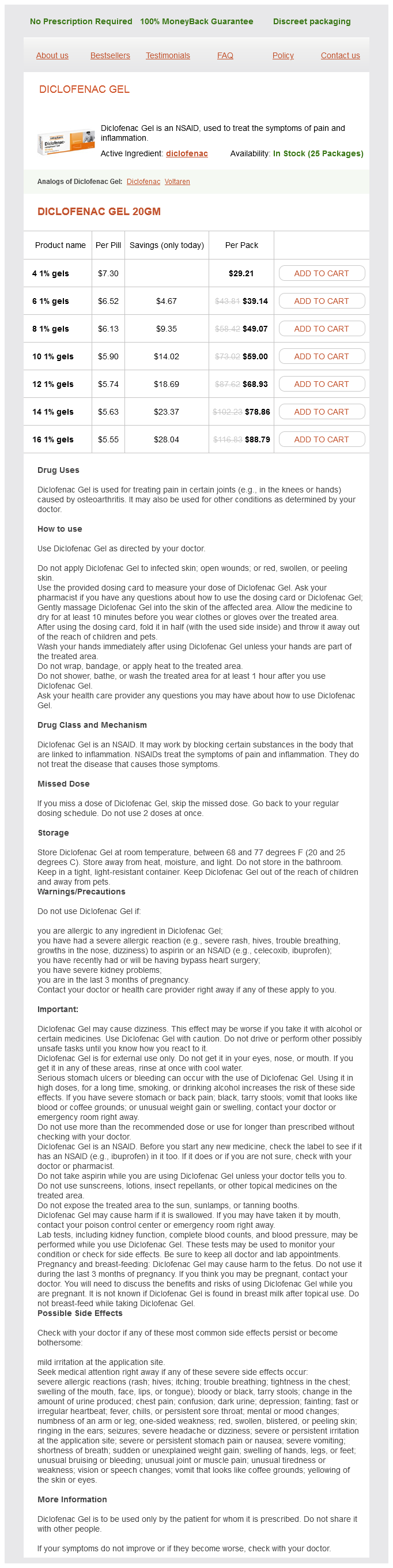
Diclofenac Gel Dosage and Price
Diclofenac Gel 20gm
- 4 1% gels - $29.21
- 6 1% gels - $39.14
- 8 1% gels - $49.07
- 10 1% gels - $59.00
- 12 1% gels - $68.93
- 14 1% gels - $78.86
- 16 1% gels - $88.79
Vasoconstriction in the inactive vascular beds contributes to the maintenance of a normal arterial blood pressure for adequate perfusion of the active tissues rheumatoid arthritis knee treatment purchase diclofenac gel 20 gm. The larger pulse pressure is primarily attributable to a greater stroke volume and, to a lesser degree, to a more rapid ejection of blood by the left ventricle. The fluid loss from the vascular compartment into contracting muscle reaches a plateau as interstitial fluid pressure rises and opposes the increased hydrostatic pressure in the capillaries of the active muscle. The fluid loss is partially offset by movement of fluid from the splanchnic regions and inactive muscle into the bloodstream. This influx of fluid occurs as a result of a decrease in hydrostatic pressure in the capillaries of these tissues and an increase in plasma osmolarity caused by movement of osmotically active particles into the blood from the contracting muscle. Severe Exercise When severe exercise is taken to the point of exhaustion, the compensatory mechanisms begin to fail. Heart rate attains a maximal level of about 180 beats/min, and stroke volume reaches a plateau. Dehydration occurs, and sympathetic vasoconstrictor activity supersedes the vasodilator influence on the cutaneous vessels. The neural activity has the hemodynamic effect of a slight increase in effective blood volume. Body temperature is normally elevated in exercise, and reduction in heat loss through cutaneous vasoconstriction can, under these conditions, lead to very high body temperatures, with associated feelings of acute distress. The reduced pH is probably the key factor that determines the maximal amount of exercise a given individual can tolerate because of muscle pain, subjective feelings of exhaustion, and inability or loss of the will to continue. The vasoconstriction produced in the inactive tissues by the sympathetic nervous system (and to some extent by the release of catecholamines from the adrenal medulla) is important for maintaining normal or increased blood pressure because sympathectomy or drug-induced block of the sympathetic adrenergic nerve fibers results in a decrease in arterial pressure (hypotension) during exercise. Some sympathetically mediated vasoconstriction occurs in active muscle and increases during strenuous exercise, when more than half of the total body musculature is contracting. In experiments in which one leg is working at maximal levels and then the other leg starts to work, blood flow decreases in the first working leg. Furthermore, blood levels of norepinephrine rise significantly in exercise, and most is derived from the sympathetic nerve endings in the active muscles. This would result in a decline in blood pressure were it not for the increasing cardiac output and constriction of arterioles in the renal, splanchnic, and other tissues. Hence the mean blood pressure Postexercise Recovery When exercise stops, sympathetic activity to the heart declines, and the heart rate and cardiac output decrease. Peripheral sympathetic activity also decreases, and coupled with resistance vessel dilation (caused by the accumulated vasodilator metabolites), arterial pressure falls, often below the preexercise level. This hypotension is brief, and the baroreceptor reflexes restore the blood pressure to normal levels. This centrally mediated (baroreceptor reflex) vasoconstriction during maximal cardiac output prevents a fall in blood pressure. This drop in blood pressure would otherwise be caused by metabolically induced vasodilation in the active muscles. If muscle O2 use were limiting, recruitment of more contracting muscle would use much more O2 to meet the enhanced O2 requirements (an amount about equal to the sum of O2 consumption of the arms and legs exercised alone). Limitation of O2 supply may be caused by inadequate oxygenation of blood in the lungs or by limitation of the supply of O2-laden blood to the muscles. Failure to fully oxygenate blood by the lungs can be excluded, because even with the most strenuous exercise at sea level, arterial blood is fully saturated with O2. Therefore O2 delivery (or blood flow because arterial blood O2 content is normal) to the active muscles appears to be the limiting factor in muscle performance. The low resting heart rate is caused by a higher vagal tone and a lower sympathetic tone. With exercise, the maximal heart rate in the trained individual is the same as that in the untrained person, but it is attained at a higher level of exercise. The trained athlete also exhibits a low vascular resistance that is inherent in the muscle. With long-term training, capillary density and the numbers of mitochondria increase, as does the activity of the oxidative enzymes in the mitochondria. After a period in which the pressure returns toward the control level, some animals continue to improve until the control pressure is attained (curve A). However, in other animals the pressure will begin to decline until death ensues (curve B). In contrast, strength exercises, such as weight lifting, appear to produce some increase in left ventricular wall thickness (hypertrophy) with little effect on ventricular chamber radius. However, this increase in wall thickness is small relative to that observed in hypertension, in which there is a persistent elevation of afterload because of the high peripheral resistance. The arterial systolic, diastolic, and pulse pressures diminish, and the arterial pulse is rapid and feeble. If sufficient blood is withdrawn rapidly from a subject to bring mean arterial pressure to about 50 mm Hg, the pressure tends to rise spontaneously toward control over the subsequent 20 or 30 minutes. In other animals (curve B), after an initial pressure rise, the pressure begins to decline, and it continues to fall at an accelerating rate until death ensues. This progressive deterioration of cardiovascular function is termed hemorrhagic shock. At some point the deterioration becomes irreversible; a lethal outcome can be retarded only temporarily by any known therapy, including massive transfusions of donor blood.

ADDII BIOTECH is proud to announce that the company's manufacturing facility located at Baddii, Himachal Pradesh has recently been approved by Food & Drug Authority of Ghana.
Our Manufacturing capability includes a wide range of therapeutic products covering almost every segment.
ADDII BIOTECH given our strong emphasis on product quality and services.