
Feldene
General Information about Feldene
In conclusion, Feldene is a widely used and efficient drug for the therapy of persistent inflammatory circumstances like rheumatoid arthritis and osteoarthritis. Its anti-inflammatory and pain-relieving properties help improve the standard of life for patients, lowering their reliance on different pain drugs. However, like all treatment, it should be taken with caution and beneath the supervision of a healthcare skilled to avoid potential unwanted effects. With correct use and monitoring, Feldene can present much-needed aid for those residing with these debilitating circumstances.
Feldene can be not suitable for everybody. Patients with a history of stomach ulcers, asthma, coronary heart or liver disease, or those that are pregnant or breastfeeding shouldn't take this treatment. It is all the time essential to debate with a well being care provider or pharmacist earlier than beginning any new medicine.
Rheumatoid arthritis (RA) is an autoimmune disorder that impacts the joints, inflicting pain, stiffness, and swelling. It is a continual condition that can lead to joint injury and disability if left untreated. Osteoarthritis (OA), then again, is a degenerative joint disease brought on by wear and tear of the joints over time. It largely affects older adults and might result in joint ache, stiffness, and decreased mobility. Both situations can considerably impact a person’s day by day activities and overall well-being.
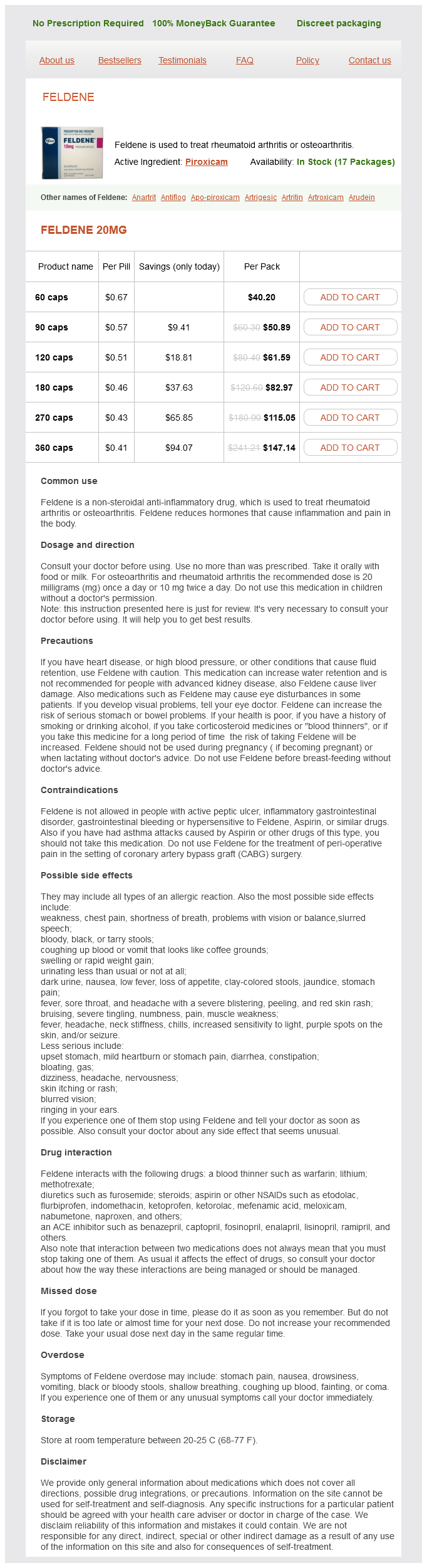
Feldene, also identified by its generic name piroxicam, is a non-steroidal anti-inflammatory drug (NSAID) used to deal with persistent inflammatory situations corresponding to rheumatoid arthritis and osteoarthritis. It belongs to the class of drugs called oxicams and works by reducing the body’s production of prostaglandins, that are answerable for inflicting inflammation, ache, and fever in the physique. Feldene might help relieve ache, stiffness, and swelling associated with these situations, permitting patients to have a greater high quality of life.
One of the benefits of Feldene over different NSAIDs is its long half-life. This signifies that the drug remains within the body for a more prolonged period, allowing sufferers to take it as quickly as a day as an alternative of multiple times a day. This can enhance affected person compliance and lower the risk of opposed unwanted effects.
However, like all medications, Feldene also has unwanted effects, the commonest being abdomen upset such as nausea, abdominal pain, and heartburn. In uncommon circumstances, it might also cause serious unwanted effects, such as an increased danger of heart assault, stroke, and stomach bleeding. Therefore, it's important to take Feldene as prescribed by the doctor and to not exceed the beneficial dose.
When taken orally, Feldene is usually prescribed at a low dose to be taken once a day. It is really helpful to be taken with food to reduce gastrointestinal unwanted facet effects, as this medication could cause stomach upset. The dose may be increased gradually if the initial dose isn't efficient, however the most really helpful every day dose mustn't exceed 20 mg. Doctors can also prescribe Feldene together with different drugs, corresponding to disease-modifying antirheumatic medication (DMARDs), to realize higher control of the disease.
Feldene helps within the management of RA and OA by decreasing the irritation and pain related to these circumstances. It is available in numerous types, together with capsules, injection, and gel. The alternative of administration is dependent upon the severity of the situation and the patient’s response to remedy.
In these cases it is the Bcells arthritis in lower back injections feldene 20 mg buy mastercard, rather than the helper Tcell, that are defective. Some immunodeficiencies can rather paradoxically cause an overactive immune response We have already mentioned that excessive production of certain classes of antibody (IgM or IgE, for example) can result from particular gene defects. It is now also clear that "immunodeficiency" affecting regulatory or tolerance mechanisms will result in an undesirable enhancement of particular types of immune response. These infants exhibit profound defects in cellular and humoral immunity and without medical intervention death occurs within the first year of life owing to severe and recurrent opportunistic infections. Prolonged diarrhea resulting from gastrointestinal infections and pneumonia due to Pneumocystis jirovecii are common; Candida albicans grows vigorously in the mouth or on the skin. If vaccinated with attenuated organisms these immunocompromised infants usually die of progressive infection. Diagnosis of primary immunodeficiencies Defects in immunoglobulins can be assessed by quantitative estimations; levels of <200 mg/dL suggest an antibody deficiency. IgG antibody responses can be measured following vaccination with tetanus toxoid, diphtheria toxoid, Haemophilus influenzae type B, and other subunit or killed vaccines but no live vaccines. Patients with Tcell deficiency will be hypo or unreactive in skin tests to such antigens as tuberculin, Candida, and mumps. The reactivity of peripheral blood mononuclear cells to the phytohemagglutinin mitogen is a good indicator of Tlymphocyte reactivity, as is also the oneway mixed lymphocyte reaction (see Chapter 15). In vitro tests for complement and for the bactericidal and other functions of neutrophils are available. If the above tests are suggestive of primary immunodeficiency, the diagnosis can be confirmed by genetic testing for mutations in the relevant gene. Gene therapy the ideal treatment for patients in which a matched transplant is not available is correction of the gene defect. The first gene therapy trials for primary immunodeficiencies were initiated over 20 years ago and there has been a steady improvement in this approach, with some setbacks along the way. A small number of patients with the Xlinked form of chronic granulomatous disease have been treated with a functional gp91phox gene but generally with only shortterm benefit. Future progress will depend upon improvements in vector design to enhance the safety and efficiency of the gene transfer, and facilitate a more precise targeting of the gene integration sites. Treatment of primary immunodeficiencies Early intervention with antibiotics and antifungals is of immediate importance, with the option of longterm lowdose prophylactic antimicrobials to prevent reinfection and subsequent complications such as hearing loss following otitis media (infection of the middle ear). Deficiencies affecting humoral responses can, to some extent, be compensated for by intravenous immunoglobulin Secondary immunodeficiency Immune responsiveness can be depressed nonspecifically by many factors. Iron deficiency is particularly important in this respect, as are zinc and selenium deficiencies. The retroviral vector containing the therapeutic gene is transfected into a packaging cell line that contains previously integrated genes encoding the essential Gag, Pol, and Env proteins. The virus particles that are produced by this cell line will lack the genes for these proteins and therefore cannot go on to produce further infectious particles following delivery of the therapeutic gene to the host hematopoi etic stem cells. Skewing of the balance between Th1 and Th2 cells as a result of infection may also depress the subset most appropriate for immune protection. Many therapeutic agents, such as Xrays, cytotoxic drugs, and corticosteroids, can have dire effects on the immune system. Another key demographic is that children under 15 years of age account for about 10% of all infected individuals. The syndrome was characterized by a predisposition to opportunistic infections. This scenario is consistent with regular direct exposure of hunters in the bushmeat trade to primate blood. The other three groups, N, O, and P, are mainly confined to Gabon, Cameroon, and neighboring countries in West Africa. The evolution of the different group M clades most probably occurred within the human population following one crossspecies transmission event. Furthermore, the discovery of a second isolate from 1960 that is highly divergent from the 1959 isolate shows that the virus had already undergone substantial diversification 50 years ago. Two evolutionary trees are shown in which the scale bar indicates 10% protein sequence divergence. Another important route of transmission is from infected mothers to their children. The chance of perinatal transmission can be significantly reduced if the mother is undergoing antiretroviral therapy. These symptoms are reminiscent of a bout of influenza and include a high spiking fever, sore throat, headaches, and swollen lymph nodes. This is referred to as the acute retroviral syndrome, the symptoms of which usually subside spontaneously in 14 weeks. Second, infected cells have an increased susceptibility to the induction of apoptosis. Third, "bystander" effects can lead to the demise of uninfected cells by exposure to viral products or molecules leading to immune activation. The plasma virus levels peak and decline to a low roughly constant level ("the set point"), which is predictive of the time of progression to disease. Pneumonia caused by the fungus Pneumocystis jirovecii is a frequent occurrence in patients and was often fatal prior to the introduction of effective antifungal therapy. For example, some viruses are naturally attenuated and are associated with slower disease progression.
In contrast arthritis in upper back discount feldene uk, Treg and memory Tcells have a lower requirement for proliferation and as such, these cells rely mainly on fatty acid oxidation for energy, with minimal dependence on glycolysis. Tcell specific metabolic signatures essential for function, and maintenance of Tcell subset, are driven by the action of key transcription factors. Damping Tcell enthusiasm We have frequently reiterated the premise that no selfrespecting organism would permit the operation of an expanding enterprise such as a proliferating Tcell population without some sensible controlling mechanisms. There are some similarities here with regulations governing corporate takeovers in the business world, where it has been deemed prudent to ensure that no single enter prise is permitted to completely dominate the marketplace. Such monopoly practices, if allowed to occur in an unregulated way, would eventually eliminate all competition. In a similar vein, in order to preserve immunological diver sity and the capacity to rapidly respond to new challenges of an infectious nature, it is necessary to ensure that Tcells specific for particular epitopes are not allowed to proliferate indefinitely and ultimately dominate the immune compartment. For these reasons, our highly adapted immune systems have evolved ways of maintaining healthy competition between Tcells, which is achieved through downregulating immune responses upon clearance of a pathogen, along with culling of the majority of recently expanded Tcells. This is also necessary because the immune compartment is of a relatively finite size and cannot accommodate an infinite number of lymphocytes. Regulatory Tcells will be discussed at length in Chapter 8, so here we will focus primarily on molecules present on activated Tcells that serve as "off switches" for such Tcells. Such molecules represent important immunological checkpoints, helping to keep Tcell responses within certain limits. Engagement of FasR on susceptible cells results in activation of the programmed cell death machinery as a result of recruitment and activation of caspase8 within the FasR complex. Active caspase8 the propagates a cascade of further caspase activation events to kill the cell via apoptosis. Therapies designed at restimulating Tcellmediated antitumor immunity are particularly desirable, as activating the adaptive immune system to target tumors not only offers an exquisite layer of precision, because of the genera tion of highly specific antigen receptors against tumor antigens, but also generates longlived memory, which may significantly lesson the chances of tumor relapse. Members of the Cbl family are protein ubiquitin ligases that can catalyze the degradation of proteins through attaching polyubiquitin chains to such molecules, thereby targeting them for destruc tion via the ubiquitinproteasome pathway. This implies that Cblb plays a major role in maintaining the requirement for signal 2 for activation of naive Tcells. Mice defective in expression of either Fas or FasL manifest a lymphoproliferative syndrome that results in autoimmune disease due to a failure to cull recently expanded lymphocytes. These and other imaging studies have revealed that the immunological synapse between the Tcell and the No. A sustained high rate of formation of successful complexes is required for full Tcell activation. Highaffinity complexes have such a long lifetime before dissociation that insufficient numbers of successful triggering events occur. Some modified peptides act as partial agonists in that they produce differential effects on the outcomes of Tcell activation. However, as we shall discuss below, certain types of antigens (called Tindependent antigens) can promote Bcell activation without the help of Tcells. The antibodies thus formed are typically of low affinity and do not undergo class switching or somatic hypermutation but provide rapid protec tion from certain microorganisms and buy time for Tdependent Bcell responses to be made. Importantly, this innatelike Bcell response is positioned at strategic areas that are sensitive to microbial invasion, such as the skin, mucosa, and the marginal zone of the spleen, where the lymphatic and circulatory systems converge. Type 1 thymusindependent antigens Certain antigens, such as bacterial lipopolysaccharides, when present at a sufficiently high concentration have the ability to activate a substantial proportion of the Bcell pool polyclonally. Scanning electron microscope analysis of a cognate Bcell/Tcell pair, embedded in 3D collagen matrix. Type 2 thymusindependent antigens Certain linear antigens that are not readily degraded in the body and that have an appropriately spaced, highly repeating determinant Pneumococcus polysaccharide, Ficoll, damino acid polymers, and polyvinylpyrrolidone, for example are also thymusindependent in their ability to stimulate Bcells directly without the need for Tcell involvement. In general, the thymusindependent antigens give rise to predominantly lowaffinity IgM responses, some IgG3 in the mouse, and relatively poor, if any, memory. Neonatal Bcells do not respond well to type 2 antigens and this has important consequences for the efficacy of carbohydrate vaccines in young children. The complex gives a sustained signal to the Bcell because of the long halflife of this type of molecule. Bcells are found in the skin and mucosal surfaces, areas con tinually under siege from pathogens, and act as a rapid first line of defense against microbial invasion. Thymusdependent antigens the need for collaboration with Thelper cells Many antigens are thymusdependent in that they provoke little or no antibody response in animals that have been thymectomized at birth and therefore have few Tcells (Milestone 7. Such antigens cannot fulfill the molecular requirements for direct stimulation: they may be univalent with respect to the specificity of each determinant; they may be readily degraded by phagocytic cells; and they may lack mitogenicity. Remember also that haptens become immunogenic when coupled to an appropriate carrier protein. Building on the knowledge that both T and Bcells are necessary for antibody responses to thymusdependent Chapter 7: Lymphocyte activation / 209 antigens (Milestone 7. Antigen processing by Bcells the need for physical linkage of hapten and carrier strongly suggests that Thelpers must recognize the carrier determinants on the responding Bcell in order to provide the relevant accessory stimulatory signals. The need for the physical union of hapten and carrier is now revealed; the hapten leads the carrier to be processed into the cell, which is programmed to make antihapten antibody and, following stimulus by the Thelperrecognizing processed carrier, it will carry out its program and ultimately produce antibodies that react with the hapten (is there no end to the wiliness of nature By carrying out these transfers with cells from animals bearing a recognizable chromosome marker (T6), it became evident that the antibodyforming cells were derived from the bone marrow inoculum, hence the nomenclature "T" for thymusderived lymphocytes and "B" for antibody forming cell precursors originating in the bone marrow. This convenient nomenclature has stuck even though bone marrow contains embryonic Tcell precursors, as the immunocompetent T and Bcells differentiate in the thymus and bone marrow, respectively (see Chapter 10). The response to tetanus toxoid in neonatally thymectomized animals could be restored by the injection of thymocytes. Different populations of cells from a normal mouse histocompatible with the recipient. They were then primed with a thymusdependent antigen such as sheep red blood cells.
Feldene Dosage and Price
Feldene 20mg
- 60 caps - $40.20
- 90 caps - $50.89
- 120 caps - $61.59
- 180 caps - $82.97
- 270 caps - $115.05
- 360 caps - $147.14
The surface of the villi is covered with a layer of epithelial cells which arthritis research back pain 20 mg feldene order, in turn, have many small projections called microvilli (collectively called the brush border) that project towards the lumen of the intestine. It has a huge surface area (about the size of a tennis court), and the chyme is forced into a circular motion as it passes through the tract, facilitating mixing and therefore digestion and absorption. Each villus contains a single, blind-ended lymphatic vessel, called a lacteal, and also a capillary network. The venous drainage from the small intestine, large intestine, pancreas and also from some parts of the stomach passes via the hepatic portal vein into the liver; here, it passes through a second capillary bed to be further processed before returning to the circulation. The small intestine absorbs water, electrolytes, carbohydrates, amino acids, minerals, fats and vitamins. The mechanisms by which movement from the lumen to the circulation occurs are variable. As the contents of the intestine are isotonic with body fluids and mostly have the same concentration of the major electrolytes, their absorption is active. Water cannot be moved directly, but follows osmotic gradients set up by the transport of ions. Both of these are against the concentration gradients, leading to a low concentration of Na+ and a high concentration of K+ within the cells. The low intracellular concentration of Na+ ensures a movement of Na+ from the intestinal contents into the cell by both membrane channels and transported protein mechanisms. Na+ is then rapidly transported out of the cell again by the basolateral Na+K+ pump. K+ leaves the cell, again via the basolateral membrane, down its concentration gradient. This outward movement of K+ is linked to an outward movement of Cl-, against its concentration gradient, Cl- having entered down its concentration gradient like Na+ via the luminal membrane. These movements set up an osmotic gradient between the lumen and the blood, leading to water absorption following the movement of Na+ and Cl- from the lumen into the cell across the luminal membrane. Carbohydrates are absorbed mostly in the form of monosaccharides (glucose, fructose and galactose). They are broken down into monosaccharides by enzymes released from the brush border (maltases, isomaltases, sucrase and lactase). The monosaccharides are transported across the epithelium into the bloodstream by means of cotransporter molecules that link Absorption of nutrients their inward movement with that of Na+ down its concentration gradient. At the basolateral membrane, monosaccharides leave the cell either by simple diffusion or by facilitated diffusion down the concentration gradient. The polypeptides produced in the stomach are broken down into oligopeptides in the small intestine by enzymes (proteases) secreted by the pancreas: trypsin and chymotrypsin. These are further broken down into amino acids by another pancreatic enzyme, carboxypeptidase, and an enzyme located on the luminal membrane epithelial cells, aminopeptidase. The free amino acids enter the epithelial cells by secondary active transport coupled to the movement of Na+ and a number of different cotransporter mechanisms. Intracellular calcium concentrations are low and any free calcium in the diet can cross the luminal membrane down a steep concentration gradient through channels or by a carrier mechanism. Most dietary iron is in the ferric (Fe3+) form which cannot be absorbed; however, in the ferrous (Fe2+) form, it forms soluble complexes with ascorbate and other substances and can be readily absorbed. These complexes are transported across the membrane by a carrier protein and, once in the cell, bind with a variety of substances including ferritin. A second carrier protein transports the iron across the basolateral membrane into the bloodstream. The major enzyme is a pancreatic enzyme called lipase which breaks fat down into monoglycerides and free fatty acids. However, before the fat can be broken down, it has to be emulsified, which is a process by which the larger lipid droplets are broken down into much smaller droplets (about 1 m in diameter). The main emulsifying agents are the bile acids, cholic acid and chenodeoxycholic acid. The free fatty acids and monoglycerides form tiny particles (45 nm in diameter) with the bile acids, called micelles. The outer region of the micelle is hydrophilic (waterattracting), whereas the inner core contains the hydrophobic (water-repelling) part of the molecule. This arrangement allows the micelles to enter the aqueous layers surrounding the microvilli, and the monoglycerides, free fatty acids, cholesterol and fat-soluble vitamins can then diffuse passively into the duodenal cells, leaving the bile salts within the lumen of the gut until they reach the ileum, where they are reabsorbed. Once within the epithelial cells, the fatty acids and monoglycerides are reassembled into fats by a number of different metabolic pathways. They then enter the lymphatic system via the lacteals and eventually reach the bloodstream through the thoracic duct. The fat-soluble vitamins, A, D, E and K, essentially follow the pathways for fat absorption. The remaining water-soluble vitamins are mainly absorbed by diffusion or mediated transport. The exception is vitamin B12, which must first bind with intrinsic factor (secreted from the parietal cells in the stomach wall). When bound, vitamin B12 attaches to specific sites on the epithelial cells in the ileum where a process of endocytosis leads to absorption. Small intestine Lower ileum 5% lost in faeces Large intestine Modi ed from Pocock G & Richards C (1999) Human Physiology: the Basis of Medicine. Pancreatic juice is made up of a number of enzymes, secreted by the acinar cells of the pancreas, which break down the major constituents in the diet.

ADDII BIOTECH is proud to announce that the company's manufacturing facility located at Baddii, Himachal Pradesh has recently been approved by Food & Drug Authority of Ghana.
Our Manufacturing capability includes a wide range of therapeutic products covering almost every segment.
ADDII BIOTECH given our strong emphasis on product quality and services.