
Glyset
General Information about Glyset
This is where Glyset is obtainable in. It belongs to a category of medicine called alpha-glucosidase inhibitors, which work by slowing down the digestion and absorption of carbohydrates within the body. Carbohydrates are the main source of sugar in our diet, so by slowing down their absorption, Glyset helps to prevent a sudden spike in blood sugar ranges after a meal. This, in flip, helps to maintain blood sugar ranges within a healthy vary.
Type 2 diabetes is a continual condition that affects hundreds of thousands of individuals worldwide. It is characterised by high blood sugar levels as a end result of body's incapability to supply enough insulin or properly use the insulin it produces. Over time, this can result in critical well being problems, corresponding to heart illness, nerve damage, and even blindness. Managing diabetes requires a combination of life-style adjustments, including a healthy diet and regular train, and sometimes treatment to control blood sugar levels.
Another good factor about Glyset is that it can be taken without regard to meals. This signifies that it doesn't should be taken earlier than or with meals like some other diabetes medicines. This added flexibility could be very helpful for individuals with busy schedules or those that wrestle with remembering to take their treatment on time.
In addition to its effectiveness in controlling blood sugar ranges, Glyset additionally has a good safety profile. It has been in the marketplace for over 20 years and has been studied in numerous clinical trials. It has also been proven to be secure for use in combination with different diabetes drugs, making it a versatile therapy option.
One of the principle advantages of Glyset is that it doesn't trigger hypoglycemia (low blood sugar) by itself. This is a typical concern for many people with diabetes, particularly those that take insulin or different medicines that can lead to low blood sugar. With Glyset, this danger is greatly reduced, making it a safer choice for a lot of people.
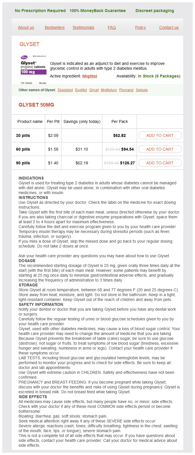
Glyset is a medication that has been making waves in the field of diabetes administration. It is an FDA-approved drug used as an adjunct to diet and exercise to improve glycemic management in adults with kind 2 diabetes mellitus. In less complicated terms, it helps to control blood sugar ranges in individuals living with diabetes, allowing them to better handle their condition and enhance their general health.
Like any treatment, Glyset does come with some potential side effects, such as bloating, diarrhea, and gasoline. These unwanted effects are normally mild and could be managed by adjusting the dosage or taking the medication with food. It is essential to debate any potential side effects with a healthcare supplier and report any regarding symptoms.
In conclusion, Glyset is a priceless medicine within the treatment of kind 2 diabetes. It offers an efficient approach to handle blood sugar levels, with out causing hypoglycemia. Its versatile dosing and good security profile make it a handy and protected choice for many people with diabetes. If you or a loved one has sort 2 diabetes, speak to your healthcare provider to see if Glyset could probably be a useful addition to your therapy plan. Remember, a mix of a healthy diet, regular exercise, and proper treatment might help you maintain higher control of your diabetes and lead a happier, more healthy life.
Clinical studies have proven that Glyset can successfully lower A1C levels in people with type 2 diabetes. A1C is a measure of average blood sugar ranges over the past 2-3 months and is used to evaluate long-term glycemic control. By reducing A1C ranges, Glyset may help to stop or delay the onset of diabetic problems, improving the general well being and high quality of life for those living with the condition.
Classification of epileptic seizures Epileptic seizures are defined as localized discount glyset 50 mg without a prescription, generalized, or unknown. Generalized seizures are conceptualized as originating at some point within, and rapidly engaging bilaterally distributed networks. The bilateral networks may include cortical and sub-cortical structures, but may not necessarily involve the whole cortex. Focal seizures are conceptualized as originating within networks limited to one hemisphere. Ictal onset is consistent from one seizure to the next with preferential propagation patterns that can involve the contralateral hemisphere. If there is uncertainty regarding classification then the seizures should be considered unclassified until further information allows accurate diagnosis. The controversy in defining whether epileptic spasms are focal or unknown has led them to remain in the unknown category. It is important to acknowledge this uncertainty particularly in children to prevent misdiagnosis. Normal or mildly abnormal results have a 100% positive predictive value at 6, 12, or 24 h of age. At 48 h of age the positive predictive value of abnormal results is 93% and negative predictive value is 71% (12). Infantile epileptic encephalopathies Epileptic encephalopathies are conditions in which cognitive, sensory, and/or motor function deterioration results from epileptic activity (14). This concept is now widely accepted as it embodies the notion that the epileptic activity itself may contribute to severe cognitive and behavioural impairments above and beyond what might be expected from the underlying aetiology alone. Included in this syndrome are: Absences with special features: · Myoclonic absence. Less specific age relationship Infancy (<1 year) Distinctive constellations Epilepsies attributed to structural: metabolic causes Childhood (112 years) Malformation of cortical development (tuberous sclerosis complex, SturgeWeber, etc. Epilepsies of unknown cause Conditions that are traditionally not diagnosed as a form of epilepsy per se. It may continue as such or evolve to partial epilepsy or severe epilepsy with multiple independent spike foci. Seizure types range from clonic seizures, myoclonic jerks, and tonic seizures, generalized tonic clonic, and focal seizures. Seizures begin with a tonic component followed by a range of autonomic and motor features, which may be unilateral, bilateral, or symmetrical. Seizures are intractable in the first few months to the first year of life with subsequent intellectual and motor impairment in the majority. It may also present with drug responsive infantile spasms with focal or lateralized epileptiform discharges. A generalized tremor appearing after the first year of life has been described (17). Proctahedrin19 this presents as an early onset epilepsy exclusively in females with variable severity, with or without mental retardation. Clinical features include early onset (mean 810months) recurrent clusters of brief seizures, fever sensitivity, tonic seizures (including focal tonic), tonicclonic seizures, focal seizures, which can generalize, intellectual disability and autistic traits. Focal seizures with ictal screaming can present in infancy within the context of hypomotor semiology or later on with prominent motor manifestations. Brainstem and cerebellar atrophy and cerebral hypomyelination are specific hallmarks. In other families Ohtahara syndrome, X- linked myoclonic epilepsy, Partington syndrome (intellectual disability with dystonic movements, ataxia, and seizures), mental retardation with tonic seizures and dystonia and infantile epileptic dyskinetic encephalopathy have also been reported. The initial component is less the 2 s, which can be followed by the second tonic component, which lasts up to 5 s. They can present soon after birth up to 5 years of age; but in the majority of children occur within the first year of life, usually between 3 and 8 months. The aetiology of this age specific syndrome is diverse ranging from Idiopathic/Genetic causes to Symptomatic/Structural brain malformations and metabolic disorders. During sleep, there is an increase in the frequency of epileptiform activity with discharges at times appearing more synchronous. Atypical hypsarryhthmia occurs when the discharges appear in synchrony and are not so chaotic or when there are intermittent periods of attenuation. Unilateral or asymmetric hypsarryhthmia is seen in structural malformations such as hemimegancephaly. Spasms can occur singly or in clusters and infantile spasms can occur without hypsarryhthmia and vice versa. The most common electrophysiological ictal correlate is an electrodecrement, consisting of generalized attenuation with superimposed fast activity or a high amplitude slow-wave burst followed by attenuation. Factors associated with a poor prognosis include age of onset, symptomatic aetiology and other seizure types. Partial migrating epilepsy in infancy formerly malignant migrating partial seizures in infancy Seizures usually begin in the first 6 months of life. A first phase can start soon after birth or even on the first day of life with sporadic seizures. Based on the origin of focal ictal discharges, semiology varies and may include lateral deviation of head and eyes, twitches of eyelids, clonic or tonic jerks of one or both limbs, apnoea, flushing and/or cyanosis of the face, chewing movements, mastication, and secondary tonic generalization.
Contribution of transcranial magnetic stimulation to the understanding of cortical mechanisms involved in motor control buy 50 mg glyset overnight delivery. A checklist for assessing the methodological quality of studies using transcranial magnetic stimulation to study the motor system: an international consensus study. Corticospinal volleys evoked by electrical stimulation of human motor cortex after withdrawal of volatile anaesthetics. Changes in the recruitment threshold of motor units produced by cutaneous stimulation in man. Role of persistent sodium and calcium currents in motoneuron firing and spasticity in chronic spinal rats. Forces consistent with plateau potentials evoked in patients with chronic spinal cord injury. Sensitivity of H-reflexes and stretch reflexes to presynaptic inhibition in humans. Evidence for further recruitment of group I fibres with high stimulus intensities when using surface electrodes in man. Correlation of the inhibitory post-synaptic potential of motoneurones with the latency and time course of inhibition of monosynaptic reflexes. Suppression of the H reflex in humans by disynaptic autogenetic inhibitory pathways activated by the test volley. H-reflex and F-responses are not equally sensitive to changes in motoneuronal excitability. Modelling recurrent discharge in the spinal motoneuron: reappraisal of the F wave. Post-activation depression of the soleus H reflex measured using threshold tracking. Transcranial magnetic stimulation and amyotrophic lateral sclerosis: pathophysiological insights. Unility of transcranial magnetic stimulation in delineating amyotrophic lateral sclerosis pathophysiology. Acquisition of a simple motor skill: task-dependent adaptation plus long-term change in the human soleus H-reflex. Operant conditioning of a spinal reflex can improve locomotion after spinal cord injury in humans. Peripheral electrical stimulation to induce cortical plasticity: a systematic review of stimulus parameters. Time course of the induction of homeostatic plasticity generated by repeated transcranial direct current stimulation of the human motor cortex. Conduction velocities of muscle and cutaneous afferents in the upper and lower limbs of human subjects. Long-latency stretch reflexes of two intrinsic muscles of the human hand analysed by cooling the arm. Relative strength of synaptic input from short-latency pathways to motor units of defined type in cat medial gastrocnemius. Jef ferys Introduction Neuronal signalling depends crucially on the movement of ions across plasma (external) membranes through various kinds of specialized ion channel. The first part of this chapter will outline cellular mechanisms responsible for cortical (and other neural) activity. Electrical (and, less commonly, magnetic) recordings provide the fastest temporal resolution for detecting neuronal activity in the brain. The distance between the recording sites on the scalp and the cortical tissue, which generates the signal, means that spatial resolution is limited, with each electrode recording from an area of several square centimetres. Higher spatial resolution can be achieved by placing electrodes onto the cortical surface. Intracortical recordings with microwires and other microscopic recording electrodes can detect the activity of single neurons or groups of neurons, although in general these are used more for research than for clinical investigation. Neurons, in contrast, contain higher concentrations of K+ and lower concentrations of Na+ than the extracellular fluid. By definition, ion channels are selective for what they allow through the otherwise impermeable membrane. They may open in response to changes in membrane potential and/or the binding of chemicals to receptor sites within the ion channel protein, or they may be constitutively open. At rest neuronal membranes are primarily permeable to K+ ions due to the presence of what are called leak channels (or more specifically, tandem pore domain K+ channels). Single cells Neurons, like all living cells, are bounded by a phospholipid membrane that prevents the free movement of chemicals, including charged ions. Electrophysiological signalling relies on the existence of gradients of ions across the cell membrane and on the controlled flow of those ions through specialized ion channel and transporter molecules. The extracellular fluid contains relatively low concentrations (~3 mM) of potassium ions (K+) and high concentrations (C) Leak K+ current Basal dendrites Axon +. K+ diffuses through the leak channel under its chemical gradient (C, top), making the inside of the neuron more negative than the outside. The resulting electrical gradient drives the positively charged K+ ions into the cell (C, bottom). The transfer of positive charge outwards results in the inside of the neuron becoming more negative than the outside. This continues until the voltage difference builds up sufficiently to cause movement of positively charged K+ ions back into the neuron at a rate that balances the movement due to the outward chemical gradient. The real resting potential of neurons (approximately -65 mV) is more depolarized than the Nernst equilibrium potential for the known difference between intracellular and extracellular K+ concentrations (of the order of -90 mV). In practice other ion channels contribute to the resting potential measured in central neurons, with notable contributions from Na+ and Cl ions.
Glyset Dosage and Price
Glyset 50mg
- 30 pills - $62.82
- 60 pills - $94.54
- 90 pills - $126.27
This is quite different from the situation when studying adults where the differentiation between peripheral nerve entrapments and multiple root entrapment makes it mandatory to sample widely order glyset on line amex. Often the changes are quite prominent and therefore the detailed examination recommended in children is not necessary. The diaphragm is easily approached, even in the youngest babies, by the technique pioneered by Bolton using a trans-costal method (46). Despite concerns from studies of ultrasound of the diaphragm (47), from anecdotal experience this appears to be a safe procedure in children, with over 50 having been performed at all ages with none having the misfortune to suffer a pneumothorax (48). The diaphragm is sampled most commonly in the intensive care ward, where a complication such as pneumothorax is perhaps less worrisome than in an outpatient setting. Combined with phrenic nerve stimulation an effective assessment of the nerve integrity to the diaphragm can be made. The blink reflex is one of these tests like the sympathetic skin response, which theoretically should have an enormous contribution to the assessment not only of the fifth and seventh cranial nerves, but also the multiple pathways, which serve it in the brainstem. The reality is otherwise; in our experience we have found it really very difficult reliably to obtain the later components of the blink reflex. Whether this is because we commonly use this technique when assessing bulbar palsy or whether this really is the situation in children is hard to determine. Of the other tests available motor unit number estimation (5153) is perfectly feasible to do in children and is well tolerated. Exercise tests also are easily tolerated by children and will be discussed in the section on myotonia. While we have no experience of nerve excitability studies (54) these should be possible to do in children, as reported by Michelle Farrar in babies (personal communication), and will feature more in the future. The structure of this chapter will follow anatomy; so start with the anterior horn cell, passing from the nerve to the neuromuscular junction, before finally considering conditions affecting the muscle. Within this anatomical description the aetiologies will follow routine assessment namely separating into congenital and acquired conditions. Within the latter, discussion will cover the different aetiologies that may be relevant from infective, autoimmune, metabolic, vascular, iatrogenic, trauma, drugs and toxins, and finally unknown causes. There are many more neuromuscular disorders, which affect children than will be discussed in this chapter. The presence of fibrillation potentials was certainly a common feature although this does not allow distinction between a myopathy and a neuropathy, but the major concern was that the established changes of chronic reinnervation often were not present. More commonly nowadays one is encountering anterior horn cell disease without any known aetiology. Also we have seen such changes in children suffering definite hypoxic ischaemic events causing cerebral palsy. Previously their weakness was explained entirely on the basis of the upper motor neuron lesion. It is important at this point to emphasize the importance of examination of the tongue in the investigation of these children. Instead in children the differential diagnosis lies between segmental involvement of the spinal cord either in the cervical or lumbar region only, or a more generalized abnormality. To this end when evidence of anterior horn cell disease is seen in tibialis anterior the next step is not exhaustively to demonstrate bilateral involvement and involvement of other segments, but to go straight away to examination of the tongue approached by the submental route. Most muscular dystrophies should not come to the attention of the electromyographer, as diagnosis is clinical, backed by genetic and muscle biopsy analysis. If the tongue is normal it is important then to identify how far up the reinnervation occurs so examination in the arm follows, which, if normal, is then followed by the examination of the contralateral leg. If only the legs have been involved a lumbar segmental involvement can be confidently diagnosed. Acquired anterior horn cell disease Segmental myelopathy has already been discussed and this may be seen particularly in the neonate where it is likely to be the result of vascular involvement of the anterior spinal arteries. The children have completely flaccid arms with preserved sensory responses, but often little or no motor response. We have seen the infarction expand further down the spinal cord to involve the thoracic region and in these circumstances the nerve supply to the intercostals is affected (67). The situation is far more serious and the children are unable to breathe without ventilation. This is a completely different prognostic situation to those with an isolated cervical involvement. The problem in United Kingdom is that the cases tend to be sporadic rather than epidemic, since the only common reason for epidemics, the polio virus, has been eliminated. However, it is really important to consider these conditions when presented with the child who has a patchy weakness particularly bulbar, following an infective condition. In this context is perhaps one of the most important recent developments in paediatric neurology, with the discovery that a proportion of children suffering Brown Vialetto van Laere disease have been proven to have a disorder of their riboflavin metabolism (8287). More remarkably treatment with large doses of riboflavin can reverse the inexorable and relentless progression of this disease previously regarded as a fatal condition. The importance of this discovery is that it is now vital to look in detail at children developing progressive weakness particularly when involved in the bulbar region in order to confirm whether there is a neurogenic abnormality. Some of the children passed very rapidly through this phase of motor neuronopathy to develop a sensorimotor neuropathy. The most well recognized cause of this phenomenon is in muscular Disorders of peripheral nerve Hereditary disorders of nerves are an ever expanding and important part of paediatric electromyography.

ADDII BIOTECH is proud to announce that the company's manufacturing facility located at Baddii, Himachal Pradesh has recently been approved by Food & Drug Authority of Ghana.
Our Manufacturing capability includes a wide range of therapeutic products covering almost every segment.
ADDII BIOTECH given our strong emphasis on product quality and services.