
Kamagra Chewable
General Information about Kamagra Chewable
In conclusion, Kamagra Chewable is a convenient and cost-effective various to Viagra for treating ED. Its delicate chewable type is easy to take, making it suitable for many who have problem swallowing tablets. It works shortly and successfully, permitting men to realize and maintain a satisfying erection. As with any medicine, it is very important consult with a physician before starting any treatment. With correct use and following the steerage of a healthcare professional, Kamagra Chewable can help males regain their sexual confidence and improve their general quality of life.
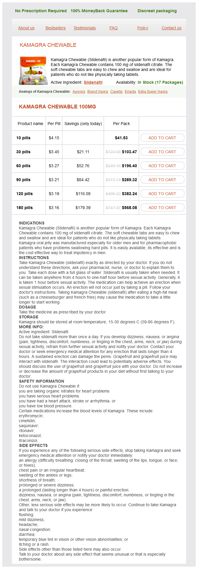
Kamagra Chewable is a drugs used to treat erectile dysfunction (ED) in men. It is a generic version of the well-known Viagra, nevertheless it is obtainable in a unique form which will attraction to those that have a tough time swallowing drugs. Kamagra Chewable is a delicate chewable pill that incorporates a hundred mg of sildenafil citrate, the lively ingredient in Viagra, and is designed to dissolve shortly within the mouth.
One of the main benefits of Kamagra Chewable is its convenience. For those that have issue swallowing drugs, the soft chewable tabs are a fantastic option. They may be easily chewed and swallowed without the need for water, making them perfect for patients of all ages. Additionally, the short dissolve characteristic of Kamagra Chewable implies that it can start working in as little as quarter-hour, compared to the 30-60 minutes it takes for Viagra to take effect.
Furthermore, Kamagra Chewable is on the market at a fraction of the value of Viagra. This is because it is a generic model and does not have the identical brand name and advertising costs related to it. Despite being more affordable, Kamagra Chewable is simply as effective as its branded counterpart. The Food and Drug Administration (FDA) has additionally accredited sildenafil citrate as secure and efficient for treating ED, making Kamagra Chewable a dependable and trusted medication for males.
The primary function of Kamagra Chewable is to increase blood circulate to the penis, permitting for a agency and lasting erection. This is achieved by blocking the enzyme known as phosphodiesterase kind 5 (PDE5), which is answerable for stopping the relaxation of the sleek muscle tissue in the penis. As a result, the blood vessels in the penis widen and allow for better blood flow, leading to an erection. It is important to notice that Kamagra Chewable doesn't act as an aphrodisiac and requires sexual stimulation to work.
It is important to notice that like any medication, Kamagra Chewable may have some unwanted side effects. These can embrace headache, facial flushing, dizziness, blurred imaginative and prescient, and indigestion. These unwanted side effects are normally mild and temporary, and if they persist or turn out to be bothersome, it is strongly recommended to consult a doctor. It can also be necessary to avoid taking Kamagra Chewable when you have certain health conditions or are taking any medications that may interact with sildenafil citrate.
It is important to examine the knee for the ability to actively extend the knee erectile dysfunction pump australia order kamagra chewable 100 mg with mastercard, since quadriceps tendon ruptures, patella fractures, or patella ligament ruptures can result in a loss of active knee extension, requiring surgery. Nondisplaced patella fractures can be treated nonoperatively with a cast or knee immobilizer, holding the knee in full extension, and weight bearing is permitted. Displaced or comminuted fractures require surgery with either tension band wiring or screws. Quadriceps tendon and patella ligament ruptures with loss of active knee extension are treated with suture repair. After surgery, the knee is held in extension and knee flexion is slowly increased over several weeks using a hinged knee brace. Patella dislocations are common injuries that occur when the femur is forcibly internally rotated on an externally rotated tibia while the foot is planted on the ground. Patients present with a significant knee effusion and on physical exam may elicit a positive apprehension test, in which a lateral force to the patella elicits pain and the sensation of an impending dislocation. Dislocated patellas can be reduced by extending the knee and manual reduction, and are treated with temporary knee immobilization. There is a high risk for recurrent dislocations, which may require surgical intervention. The danger however is due to the close proximity of the popliteal artery that runs directly behind the knee, which may kink or suffer a tear of the intimal wall when the knee dislocates. A neurovascular exam is extremely important, followed by immediate reduction of the knee and repeat exam of the pulses. If there is evidence of diminished or absent pulses, an angiogram must be performed, and vascular surgery may need to perform emergent vascular repair. With regard to the the tibial plateau is comprised of the articular surfaces and underlying cancellous bone of the medial and lateral plateaus of the proximal tibia. Fractures of the plateau result from axial loads sustained in falls from a height or high energy trauma, and are often associated with injuries to the menisci and cartilage of the knee. Fractures can involve the medial, lateral, or both plateaus with significant comminution, angulation, and depression, creating a challenging injury to fix. Minimally displaced fractures may be treated nonoperatively with strict nonweight bearing until the fracture heals. Fractures associated with displaced articular fragments require surgery in order to restore the smooth contour of the articular surface. Since there is often a depression of the cancellous bone, bone graft or bone substitutes may be needed to buttress the articular surface and restore the anatomic alignment of the tibia. Patients are kept strictly nonweight bearing for several weeks until the fracture begins to heal, though early range of motion is encouraged. Repair of ligament or meniscus injuries may also be indicated at the time of surgery. Trauma and direct blows to the tibia result in transverse or comminuted fracture patterns, while torsional injuries cause spiral fractures. Fractures with minimal angulation can be treated with reduction and casting, followed by transition to a functional brace and slow return to weight bearing, and may need to be immobilized for several months since these fractures can be slow to heal. Most tibial shaft fractures, especially comminuted and angulated fractures, are treated with an intramedullary nail placed down the tibial canal, with interlocking screws placed proximally and distally, and weight bearing can begin soon after surgery. Plate and screw fixation can also be used, however since the tibia is subcutaneous, hardware placed along the shaft can increase the risk of wound breakdown, and therefore intramedullary nailing is the preferred treatment. Fibula shaft fractures often occur along with tibial shaft fractures, though they usually heal well without surgery. Tibial Plafond (Pilon) Fractures the tibial plafond is the distal tibial articular surface of the ankle joint. Pilon fractures are typically high energy injuries from axial compression or a shear force. These injuries can cause significant soft tissue injury, severely comminuted intra-articular fragments, and wound healing problems, making these fractures very difficult to treat. Due to the soft tissue injury, these fractures are initially treated with external fixation until the swelling subsides, which may take several days to weeks. The goals of surgery are to restore the articular surface, fix the fibula in order to maintain and establish anatomic length, bone graft any cancellous bone defects, and stabilize the distal tibia with plate and screw fixation. Despite best efforts, patients may suffer from ankle pain and stiffness, arthritis, wound healing problems, infection, nonunion, and some patients eventually need ankle fusion in the future. Lateral Malleolus Fractures Isolated fractures of the lateral malleolus require anatomic reduction of the fracture in order to restore normal ankle joint congruity. The talus can sublux laterally following lateral malleolus fractures, and even 1 millimeter of talar shift decreases the surface contact between the talus and the tibia by 40%, increasing the risk of developing arthritis. Closed reduction and casting can be successful, however if the fracture cannot be adequately reduced, then open reduction internal fixation of the fibula is done with plate and screw fixation. Medial Malleolar Fractures An isolated fracture of the medial malleolus is usually an avulsion-type injury. Minimally displaced fractures can be treated with a cast or walking boot, while displaced fractures are fixed with screws placed up through the tip of the malleolus. Bimalleolar Fractures Fractures to both the medial and lateral malleoli often require surgery. These injuries are more unstable and the talus will often sublux or completely dislocate laterally. Occasionally, the posterior articular surface of the distal tibia, or posterior malleolus, can be fractured as well, resulting in a trimalleolar ankle fracture. Often it is a small fragment and does not need to be fixed, however if it involves >25% of the articular surface it should be fixed with screws placed either anteriorly or posteriorly. Syndesmosis injuries the syndesmosis is comprised of several ligaments between the distal tibia and fibula that provide stability to the ankle joint by resisting axial, rotational, and translational forces. The syndesmosis can be disrupted at the time of ankle fractures and requires special attention.
Much is known about the progression of these structures from their normal state (called bands) to their contracted state (called cords) erectile dysfunction at age 27 buy discount kamagra chewable 100 mg line, but little is known on how or why this process begins. These collagen bundles, with the aid of myofibroblasts, contract down to form the cords, which are the hallmark of the symptomatic patient. Corticosteroid injections may soften nodules and decrease the discomfort associated with them but are ineffective against cords. Long-term studies have demonstrated more rapid recovery from needle fasciotomy, as the procedure is called, but more durable results with fasciectomy. Great care must be taken to preserve as much of the subdermal vascular plexus with the elevated skin flaps to minimize postoperative skin necrosis. All nerves, tendons, and blood vessels in the operative field should be identified. Once this is done, the involved cord is resected while keeping the critical deeper structures under direct vision. Alternative cord resection techniques include removal of the skin over the contracture (dermatofasciectomy). This requires a skin graft to the wound and should only be done if skin cannot be separated from the cords and local tissue cannot be rearranged with local flaps to provide closure of the wound. Digital nerve injury can be quite devastating, producing annoying numbness at best or a painful neuroma in worse situations. Hand therapy is typically instituted within a week of surgery to begin mobilization of the fingers and edema control. The therapist can also identify any early wound problems because he or she will see the patient more frequently than the surgeon. Extension hand splinting is maintained for 4 to 6 weeks, with nighttime splinting continued for an additional 6 to 8 weeks. After this point, the patient is serially followed for evidence of recurrence or extension of disease. Other predisposing factors include diabetes, neuropathies, and immunocompromised patients. Proper treatment consists of incision and drainage of any collections followed by débridement, obtaining wound cultures, antibiotic therapy, elevation, and immobilization. Staphylococcus and Streptococcus are the offending pathogens in about 90% of hand infections. Infections caused by intravenous drug use or human bites and those associated with diabetes will often be polymicrobial, including gram-positive and gram-negative species. Although -hemolytic Streptococcus and Staphylococcus aureus are the most commonly encountered pathogens in human bites, Eikenella corrodens is isolated in up to one-third of cases and should be considered when choosing antimicrobial therapy. Ziehl-Neelsen staining and cultures at 28°C to 32°C in Lowenstein-Jensen medium must be performed if there is a suspicion for atypical mycobacteria. Group A -hemolytic Streptococcus is the most common offending pathogen and causes a more diffuse spread of infection. Septic arthritis, osteomyelitis, an abscess, a deep-space infection, and necrotizing fasciitis are severe infectious processes that may initially mimic cellulitis. These must be ruled out appropriately before initiating treatment, and serial exams should be conducted to ensure proper diagnosis. Treatment of cellulitis consists of elevation, splint immobilization, and antibiotics that cover both Streptococcus and Staphylococcus. Failure to improve after 24 hours indicates a need to search for an underlying abscess or other infectious cause. Treatment consists of incision and drainage with appropriate débridement, wound cultures, wound packing, elevation, immobilization, and antibiotics. The packing should be removed in 12 to 24 hours or sooner if there is clinical concern, and warm soapy water soaks with fresh packing should be initiated. Delayed primary closure should only be performed after repeat washouts for larger wounds where complete infection has been achieved. Postoperatively, the patient went on to heal all his incisions and, with the aid of weeks of hand therapy, recover full motion. Collar-Button Abscess this is a subfascial infection of a web space and is usually caused by skin trauma that becomes infected; it often occurs in laborers. The adherence of the palmar web space skin to the palmar fascia prevents lateral spread, so the infection courses dorsally, resulting in both palmar web space tenderness and dorsal web space swelling and tenderness. Incision and drainage, often using separate volar and dorsal incisions, is mandatory, and follows the same treatment as for any abscess or deep-space infection. Osteomyelitis Osteomyelitis in the hand usually occurs due to an open fracture with significant soft tissue injury. The presence of infected hardware, peripheral vascular disease, diabetes, and alcohol or drug abuse are also predisposing factors. Presentation includes persistent or recurrent swelling with pain, erythema, and possible drainage. It will take 2 to 3 weeks for periosteal reaction and osteopenia to be detected on radiographs. Treatment consists of antibiotics alone in the early stage as long as there is favorable response. Antibiotic therapy is continued for 4 to 6 weeks once the patient clinically improves and there is no further need for débridement. For osteomyelitis in the setting of an acute fracture with internal fixation in place, the hardware should erythema, and tenderness. Radiographs may show a foreign body or fracture, with widened joint space early in the process and decreased joint space late in the process due to destruction. Joint aspiration with cell count, Gram stain, and culture is used to secure the diagnosis.
Kamagra Chewable Dosage and Price
Kamagra Chewable 100mg
- 10 pills - $41.53
- 30 pills - $103.47
- 60 pills - $196.40
- 90 pills - $289.32
- 120 pills - $382.24
- 180 pills - $568.08
Capillary refill erectile dysfunction humor cheap kamagra chewable uk, turgor, Doppler signal, and bleeding to pinprick all provide useful information regarding vascular status. The finger or hand with vascular compromise requires urgent operative exploration. Unlike the complete amputation, in which the amputated part can be cold preserved (see below), devascularization without amputation produces warm ischemia, which is tolerated only for a matter of hours. At least one of the palmar arterial arches is intact in 97% of patients,5 so this will usually not compromise hand perfusion. Each digit also has two arterial inflows and can survive on one (see Amputations and Replantation section below). In the academic hospital setting, however, consideration should be given to repairing all vascular injuries. Instructing a resident in vascular repair in the noncritical setting will produce a more skilled and prepared resident for when a critical vascular injury does arise. Skin may be closed with permanent or absorbable sutures; absorbable sutures will spare the patient the discomfort of suture removal several weeks later. For more proximal unsalvageable amputations, revision should be performed in the operating room to maximize vascular and neural control. The more proximal the amputation, the more important to function the prosthesis is likely to be. Flexor digitorum superficialis insertion to the flexor digitorum profundus insertion. Over the ensuing decades, more stringent guidelines have been established regarding what should be replanted. Indications for replantation include amputations of the thumb, multiple digit amputations, and amputations in children. As with all guidelines, one should evaluate the particular needs of the injured patient. In preparation for replantation, the amputated part and proximal stump should be appropriately treated. The amputated part should be wrapped in moistened gauze and placed in a sealed plastic bag. Do not use dry ice, and do not allow the part to contact ice directly; frostbite can occur in the amputated part, which will decrease its chance of survival after replantation. Bleeding should be controlled in the proximal stump by as minimal a means necessary, and the stump should be dressed with a nonadherent gauze and bulky dressing. The usual history is that a door closed on the finger (commonly the middle, due to its increased length) or something heavy fell on the finger. Initial evaluation should include: wound(s) including the nail bed, perfusion, sensation, and presence and severity of fractures. To properly assess the nail bed, the nail plate (hard part of the nail) should be removed. Great care must be taken when suturing because excessive traction with the needle can further lacerate the tissue. This is done to prevent scarring from the nail folds down to the nail bed that would further compromise healing of the nail. In some situations, tissue may have been avulsed in the injury and be unavailable for repair. For wounds less than 1 cm2 with no exposed bone, secondary intention will produce excellent functional and aesthetic results. For larger wounds or wounds or with bone exposed, one must decide if the finger is worth preserving at the current length or if shortening to allow for primary closure is a better solution. A useful guideline is the amount of fingernail still present; if greater than 50% is present, local or regional flap coverage may be a good solution. A wide range of antegrade and retrograde homodigital flaps can be mobilized to cover the defect. Some carry sensation or can receive nerve coaptation to recover sensation over time. For wounds too large to cover with homodigital tissue, regional flaps can be considered. The finger is maintained in flexion for 14 to 21 days until division of the flap pedicle and inset of the flap. Some authors have reported prolonged stiffness in patients over 30 years old, but careful flap design helps minimize this complication. High-pressure devices are commonly used for cleaning and applications of liquids such as lubricants and paint. Most commonly, the inexperienced worker accidentally discharges the device into his nondominant hand at the base of the digit. Severity of injury depends on the amount and type of liquid injected; hydrophobic compounds cause greater damage. The patient should be informed of the severity of the injury, and exploration is ideally performed within 6 hours of injury. Up to 50% of such injuries result in loss of the digit, but early recognition and treatment are associated with increased chance of digit survival. On the dorsum of the forearm, there is the dorsal compartment as well as the mobile wad compartment, beginning proximally over the lateral epicondyle. Extrinsic causes include splints and dressings that are circumferentially too tight and intravenous infiltrations. Infiltrations with hyperosmolar fluids such as x-ray contrast are particularly dangerous, because additional water will be drawn in to neutralize the hyperosmolarity. Measurement of compartment pressures can be a useful adjunct to assessment of the patient. The Stryker pressure measurement device or similar device is kept in many operating rooms for this purpose.

ADDII BIOTECH is proud to announce that the company's manufacturing facility located at Baddii, Himachal Pradesh has recently been approved by Food & Drug Authority of Ghana.
Our Manufacturing capability includes a wide range of therapeutic products covering almost every segment.
ADDII BIOTECH given our strong emphasis on product quality and services.