
Lady era
General Information about Lady era
Another benefit of Lady era is that it's comparatively safe to make use of. It has been approved by the Food and Drug Administration (FDA) for the therapy of hypoactive sexual desire dysfunction (HSDD) in premenopausal girls. This is a condition characterized by an absence of sexual want or curiosity. By rising blood move to the clitoris, Lady period may help to fight this concern and improve sexual satisfaction for these ladies.
Some critics argue that Lady era solely focuses on physical pleasure and ignores the emotional and psychological aspects of female sexuality. However, many research have shown that sexual satisfaction for girls is carefully tied to physical pleasure. By offering intense sexual pleasure, Lady period can even enhance a lady's general sexual satisfaction and shallowness.
One of the main advantages of Lady era is that it has been specifically designed for ladies. In the previous, girls who skilled sexual dysfunction or had trouble reaching orgasm were usually prescribed drugs meant for males. This led to ineffective results and even potential side effects. With Lady period, ladies can relaxation assured that this medication has been tailor-made to their particular needs and needs.
Lady period works by concentrating on the blood move to the genital space, specifically the clitoris. In ladies, the clitoris is a small, however extremely sensitive organ that's answerable for sexual pleasure. When blood move is elevated to this area, it might possibly lead to heightened arousal, sensitivity, and finally, an intense orgasm.
This medication is not meant to be taken as a every day medication, like many male enhancement medication. Instead, it is meant to be taken on an as-needed foundation, earlier than sexual activity. This permits women to have management over after they need to expertise heightened sexual pleasure.
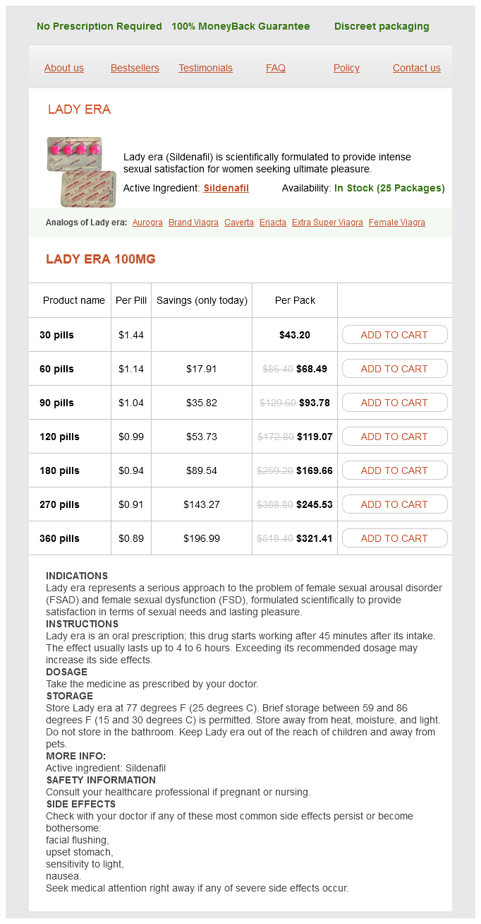
In current years, the subject of feminine sexuality and sexual satisfaction has been dropped at the forefront of many discussions. With the success of medications like Viagra for men, it was solely a matter of time before a similar product was created for women. Enter Lady era (Sildenafil), a scientifically formulated medicine that goals to supply intense sexual satisfaction for women looking for ultimate pleasure.
In conclusion, Lady era (Sildenafil) is a scientifically formulated treatment that gives intense sexual satisfaction for ladies seeking final pleasure. With its capacity to target the blood flow to the genital space, it can enhance arousal, sensitivity, and lead to highly effective orgasms. It has been particularly designed for girls, making it a protected and efficient possibility for those seeking to enhance their sexual satisfaction. With minimal side effects and its FDA approval, Lady era is quickly turning into a go-to treatment for ladies trying to enhance their sexual experience.
Furthermore, Lady period has been proven to have minimal unwanted aspect effects. Unlike male enhancement medication that may typically trigger complications, dizziness, and flushing, the potential unwanted effects of Lady period are gentle and uncommon. These may include nausea or a mild headache, but they typically subside rapidly.
Myelinated nerve fibers breast cancer questions for doctor purchase 100 mg lady era free shipping, which are sensory, also supply adventitia of blood vessels. This narrow, thin-walled vessel (arrows) has a uniform caliber, and its lumen contains a row of lymphocytes. Smooth muscle cells make up the arteriole wall; erythrocytes are in the lumen of the venule. A wide intercellular gap (circle) separates ends of two endothelial cells of the lymphatic. The thicker endothelium of the blood capillary is sectioned at the level of a nucleus (Nu). The process of a fibroblast (Fi) is in interstitial connective tissue between the two vessels. They absorb lymph, which is a protein-rich exudate of blood, as well as electrolytes and water, from blood capillaries, with lymph being moved mainly by contraction of surrounding skeletal muscles. This fluid normally fills extracellular connective tissue spaces, and some is reabsorbed back into the venous end of blood capillaries. Lymphatic capillaries continually take up excess fluid, plus wandering lymphocytes and other cells, and eventually add them back to the systemic circulation. They thus play a role in homeostasis by regulating interstitial fluid pressure and maintaining plasma volume. Each day, lymphatics return about 40% of total plasma protein to veins and permit entry of chylomicrons and immuno- 8. Lymphatics also remove foreign substances from tissues and aid in clearance of debris after tissue injury. They consist of one layer of flat endothelial cells, which are thinner and slightly larger than those of blood capillaries, but no pericytes. An absent or incomplete basal lamina facilitates permeability of these vessels to large molecules and cells. The endothelium features transcytotic vesicles, which engage in transport, and actin filaments, which are contractile. Lymphatic capillaries coalesce into larger vessels that resemble veins and transport lymph via large lymphatic channels and ducts to the venous circulation. Like veins, lymphatic channels have three concentric tunics, although they are not as clearly delineated as they are in veins of similar size. The duct appears slightly collapsed in section; its wall is thin relative to the size of the lumen. Loose connective tissue of the adventitia contains irregularly arranged collagen (Co) and many vasa vasorum. Although their structure is similar to that of veins of comparable size, the three concentric tunics are not as clearly demarcated. It conveys a mixture of lymph and chyle (emulsified fats and free fatty acids) from the cisterna chyli (an elongate, lobulated sac) in the abdomen and upward through the thorax to the left subclavian vein in the root of the neck. The tunica intima of the thoracic duct consists of endothelium, delicate subendothelial connective tissue, an inconspicuous internal elastic lamina, and some longitudinal smooth muscle. The media is the thickest layer and has a mixture of circular and helically arranged smooth muscle that intermingles with a rich network of elastic fibers. The tunica adventitia is poorly defined and blends with adjacent loose connective tissue. It has an extensive network of vasa vasorum that extends into the outer layer of the media and may also contain a few bundles of longitudinal smooth muscle. Changes in pressure in the body associated with breathing assist movement of lymph along the thoracic duct, which is also provided with many valves to prevent fluid backflow. Arranged in pairs, they consist of a folding of endothelium with a thin collagenous connective tissue core. Just before the thoracic duct enters the subclavian vein, it is joined by the left jugular and subclavian trunks, which carry lymph from the left side of the head and neck and the left arm. The most common causes of this life-threatening condition are complications from malignant lymphoma and trauma caused by thoracic, mediastinal, or cardiac surgery. Treatment options include pleural drainage; thoracic duct ligation via thoracoscopy; omitting fat from the diet; and treatment with noninvasive subcutaneous medications, such as synthetic analogues of somatostatin, to prevent fluid effusion. Lymphatics of upper limb Tonsils Three-dimensional view of jejunal wall Organization of lymphatic system. This tissue is located in the appendix, so it is more accurately called gut-associated lymphoid tissue. Its major functions are thus to serve as a source of immunocompetent cells that can react with and neutralize antigens and to distinguish self from nonself. The system comprises lymphoid tissues and organs whose main constituents are aggregates of lymphocytes and other cells of the mononuclear phagocyte system. These cells are enmeshed in a supportive framework (stroma) of reticular cells and fibers, so lymphoid tissue is classified as a specialized reticular connective tissue. More densely packed, spherical clusters of lymphocytes called lymphoid nodules (or follicles) may also be found in these and other sites. The nodules may appear as single collections of lymphocytes or as more permanent, multiple aggregates, such as tonsils and Peyer patches. Discrete lymphoid organs may be encapsulated (lymph nodes, thymus, and spleen) or unencapsulated (bone marrow).
The cells phagocytose antigen-antibody complexes and parasites women's health clinic barrie lady era 100 mg purchase line, and elevated cell numbers occur in parasitic infections and allergic responses such as hay fever and asthma. Eosinophils play a central role in controlling parasitic diseases such as schistosomiasis. They kill parasitic larval helminths by releasing toxic molecules from the specific granules. In bronchial asthma and allergic eczema, local eosinophil accumulations in tissues may cause severe cell injury and necrosis. These harmful effects are likely related to major basic protein and eosinophil cationic protein, which are components of the specific granules. The easily recognized basophil (Ba) has many large basophilic specific granules that are blue. These granulocytes-between neutrophils and eosinophils in size, with a diameter of 10-14 mm-have large, distinctive specific granules that are intensely basophilic and fill the cytoplasm. In blood smears, the nucleus is usually obscured by many closely packed basophilic granules, which often stain more deeply than nuclear chromatin. Basophils closely resemble mast cells of connective tissue; their granules, like those of mast cells but larger and fewer, have metachromatic staining properties and contain histamine and heparin. Basophils also produce platelet-activating and eosinophilic chemotactic factors, which exert powerful pharmacologic effects outside the circulation. Basophils probably originate in bone marrow from cell precursors different from those of mast cells. In both cell types, histamine released by exocytosis at cell surfaces increases vascular permeability during an inflammatory response. Both cells also have surface receptors that bind immunoglobulin IgE, which is produced by plasma cells in connective tissue. The motile basophils are involved in allergic reactions and increase in number in many clinical conditions such as hay fever, urticaria, chronic sinusitis, and some leukemias. Mild basophilia may be part of a general inflammatory response to some infections, for example, smallpox, chickenpox, or influenza. It also occurs in allergic disorders or autoimmune inflammation such as rheumatoid arthritis or ulcerative colitis. More often, and for unclear reasons, basophilia is noted in malignant hematologic conditions called myeloproliferative disorders. The most common such condition is chronic myeloid leukemia, in which basophils are often markedly increased, as are eosinophils, neutrophils, and immature neutrophilic forms such as band cells, metamyelocytes, myelocytes, and promyelocytes. In patients with chronic myeloid leukemia, an increasing basophil count can suggest worsening of disease. A rim of bluegray cytoplasm caps a darkly stained round nucleus in a large lymphocyte (Ly). The spherical, slightly indented nucleus contains condensed heterochromatin with patches of euchromatin. They can be classified as small (610 mm) and medium to large (11-16 mm), with most circulating lymphocytes in normal blood being small. These spherical cells have a densely stained nucleus and a thin rim of blue-gray cytoplasm. All lymphocytes derive from bone marrow stem cells; those that differentiate and mature in the thymus are T cells, and those that develop in bone marrow where they acquire specific cell surface antigens are B cells. In normal peripheral blood, 60%-80% of lymphocytes are T cells, 10%-15% are B cells, and the rest are null cells, which lack both B and T cell markers. B cells express various cell surface markers; when the cells are activated by antigen, they differentiate into plasma cells, which synthesize and 7. Cell-mediated immunity involves T cells, whereas B cells function in humoral (antibody) immunity. A small Golgi complex, small numbers of mitochondria, and occasional lysosomes are also present. It often occurs in infants and adolescents during infections that would likely produce a neutrophil response in adults. Most patients need only symptomatic treatment, as recovery is usually 4-6 weeks after symptoms begin. A monocyte (Mo) nucleus is highly indented and less densely stained than that of lymphocytes. Throughout the light blue cytoplasm are many faintly stained granules, so the cytoplasm looks dusty. The cell attaches to the endothelium (En) by pseudopodia (arrows) as it begins migrating from the lumen on its way to becoming a macrophage. This migratory process- diapedesis-enables leukocytes to leave the circulation for functions in surrounding tissues. They constitute 3%-10% of the total leukocyte count and, with a diameter of 1220 mm, are the largest leukocytes in blood smears. They usually circulate in the bloodstream for only 1-3 days and perform almost all functions outside the circulation. These actively motile cells enter connective tissues to become macrophages (or phagocytes). Each cell has a nucleus that varies in form and may have an oval, kidney, or horseshoe shape. In contrast to the coarse, dark-staining nuclear chromatin of lymphocytes, monocyte nuclear chromatin is finely granular and pale stained, and indented. Monocyte cytoplasm has a blue-gray tinge and contains a moderate number of small, scattered azurophilic granules but no specific granules. A well-formed Golgi complex is near the indentation of the nucleus; the cytoplasm also contains scattered 7. Cytoplasmic filaments and many irregular pseudopodia are typical of cell surfaces, consistent with motility.
Lady era Dosage and Price
Lady era 100mg
- 30 pills - $43.20
- 60 pills - $68.49
- 90 pills - $93.78
- 120 pills - $119.07
- 180 pills - $169.66
- 270 pills - $245.53
- 360 pills - $321.41
As more power and less fine control is needed menopause uti cheap 100 mg lady era with amex, the larger motor units are progressively recruited. The mechanical properties of the muscle fibers are also matched to the size of their motor unit. Because the muscle fibers of the smallest motor units are those most often activated, they must be relatively resistant to fatigue (see Plate 2-15). The terminals, although unmyelinated, are invested by a Schwann cell, which projects finger-like processes between the membranes of the nerve and the muscle. The nerve terminal lies in a trough within the muscle fiber membrane (sarcolemma); it is rich in mitochondria and contains numerous synaptic vesicles about 50 nm in diameter. These vesicles, which contain the neurotransmitter acetylcholine, are clustered around nipple-shaped active zones located at regular intervals along the terminal membrane. Acetylcholine is released by exocytosis of vesicles lying adjacent to both sides of the active zone. The presynaptic and postsynaptic membranes are separated by a space approximately 50 nm wide. Freeze-fracture electron microscopy reveals granular structures embedded in the postsynaptic membrane. These structures, which are concentrated on the banks of the junctional folds opposite the sites of acetylcholine release, are acetylcholine receptors that mediate the action of the transmitter. They are sparse in regions of the sarcolemma not close to the neuromuscular junction. The muscle fiber is surrounded by a connective tissue basement membrane that continues into the synaptic cleft, sending extensions into the junctional folds. It also contains other important molecules that help guide the growth of the nerve terminal during development and regeneration, determine the locations of the presynaptic active zones, and induce the clumping of acetylcholine receptors opposite the synaptic vesicle. The membrane of the nerve terminal has a different assortment of ion channels: fewer sodium channels, several types of potassium channels, and, most important, voltage-dependent calcium channels. When an action potential arrives at the nerve terminal, it opens the calcium channels and calcium ions move from the extracellular fluid, where the concentration is about 2. The sudden increase in intraterminal concentration of calcium ions is linked to the release of acetylcholine by exocytosis at the synaptic vesicle release sites. The acetylcholine binds to receptor molecules on the postsynaptic membrane, opening channels that permit the influx of sodium ions and efflux of potassium ions. The net effect is a depolarization of the muscle membrane and a triggering of the muscle action potential. The choline is conserved by active uptake into the nerve terminal, where it is reconverted into acetylcholine by the enzyme choline acetyltransferase. Organic calcium channel blockers, such as verapamil, and divalent cations, such as lead, prevent calcium ions from entering the nerve terminal and thus block release of the neurotransmitter acetylcholine. The neurotoxin botulin blocks acetylcholine release more directly by a mechanism that is still unknown. Some drugs block the acetylcholine receptors either reversibly (curare) or irreversibly (-bungarotoxin). Succinylcholine and decamethonium produce muscle relaxation not by preventing the opening of the acetylcholine-activated channels but by keeping them open too long. The voltage-dependent sodium channel, which mediates the muscle action potential, is also time dependent: prolonged depolarization inactivates the action potential. By keeping the acetylcholine channel open, the just-mentioned depolarizing blockers keep the muscle membrane depolarized and refractory to impulse initiation. Hemicholinium weakens neuromuscular transmission by blocking the reuptake of choline, thus reducing the synthesis of acetylcholine. Acetylcholine agonists, such as nicotine, react directly with the acetylcholine receptor. Others, such as physostigmine, pyridostigmine bromide, and edrophonium chloride, inhibit acetylcholinesterase and strengthen transmission by delaying the breakdown of acetylcholine. The action of physostigmine and pyridostigmine bromide persists for hours, and these agents are used to treat the neuromuscular disease myasthenia gravis; edrophonium chloride, on the other hand, is short acting and is used in diagnosing the disease. Thick filament compressed between Z bands; thin filaments interfere with one another. The amount of transmitter released at the nerve terminal of the skeletal neuromuscular junction is sufficient to produce an end plate potential that always reaches the threshold required to activate a muscle action potential. This is not the case in certain pathologic conditions, such as myasthenia gravis or the Lambert-Eaton (myasthenic) syndrome. The muscle action potential is propagated along the entire length of the muscle fiber and deep into the fiber through the transverse (T) tubules. Calcium ions are released almost synchronously from the entire sarcoplasmic reticular system, which elicits a contraction, or twitch. The tension of the twitch can be measured under conditions in which the muscle is not allowed to shorten (isometric contraction). Because all the sarcomeres are activated, the single-twitch strength of a fiber tends to be the same each time, providing the muscle remains the same length. If a second twitch is elicited before the first has relaxed, the maximum tension achieved is increased. If the muscle is activated at a high enough frequency, the twitches fuse into a continuous smooth contraction (tetanus) of even greater tension. The tension developed by a tetanically stimulated muscle depends on the final length the muscle fibers are permitted to reach. If the muscle is contracted too far, the thin filaments overlap, which interferes with their interactions with the thick filaments, reducing the maximum attainable tension. On the other hand, if the muscle is stretched, the thin filaments do not have access to all of the available myosin head groups and fewer than the maximum number of crossbridges are formed.

ADDII BIOTECH is proud to announce that the company's manufacturing facility located at Baddii, Himachal Pradesh has recently been approved by Food & Drug Authority of Ghana.
Our Manufacturing capability includes a wide range of therapeutic products covering almost every segment.
ADDII BIOTECH given our strong emphasis on product quality and services.