
Lansoprazole
General Information about Lansoprazole
The abdomen naturally produces acid to assist within the digestion of meals. However, when too much acid is produced, it might possibly lead to various well being points. Stomach ulcers, for example, are sores that develop on the liner of the stomach and may trigger burning pain and discomfort. If left untreated, they will result in critical complications similar to bleeding and perforation of the abdomen wall. Prevacid works by inhibiting the production of acid, offering reduction and allowing the ulcers to heal.
Prevacid is out there in various types, together with capsules, orally disintegrating tablets (ODT), and oral suspension. It is normally taken as quickly as a day, earlier than a meal, and should be swallowed whole for capsules and ODT, or blended with applesauce for ODTs. The oral suspension may be taken alone or blended with water earlier than use. It is essential to follow the dosage and instructions supplied by a healthcare skilled.
Prevacid can also interact with different medications, particularly blood thinners and certain antibiotics and antidepressants. It is essential to inform a healthcare skilled of all current medications earlier than starting Prevacid to avoid any potential interactions.
In conclusion, Prevacid, or lansoprazole, is a widely prescribed treatment for the treatment of stomach and intestinal ulcers, erosive esophagitis, and other circumstances involving excessive stomach acid. By decreasing the amount of acid produced in the stomach, it supplies reduction from signs and promotes healing. It is essential to take Prevacid as directed and to consult a physician when you experience any unwanted aspect effects or have any issues.
Another frequent situation that's handled with Prevacid is erosive esophagitis, a condition by which abdomen acid damages the lining of the esophagus, causing irritation and discomfort. This can happen because of acid reflux, a situation in which abdomen acid flows again up into the esophagus, causing a burning sensation within the chest, generally often identified as heartburn. Prevacid helps to decrease the quantity of acid within the abdomen, reducing the probability of acid reflux and permitting the esophagus to heal.
As with any medication, Prevacid may cause side effects. These can include headache, nausea, diarrhea, and belly pain. More severe side effects may happen in rare instances, similar to allergic reactions or increased risk of bone fractures. It is important to debate any potential side effects with a health care provider before starting Prevacid.
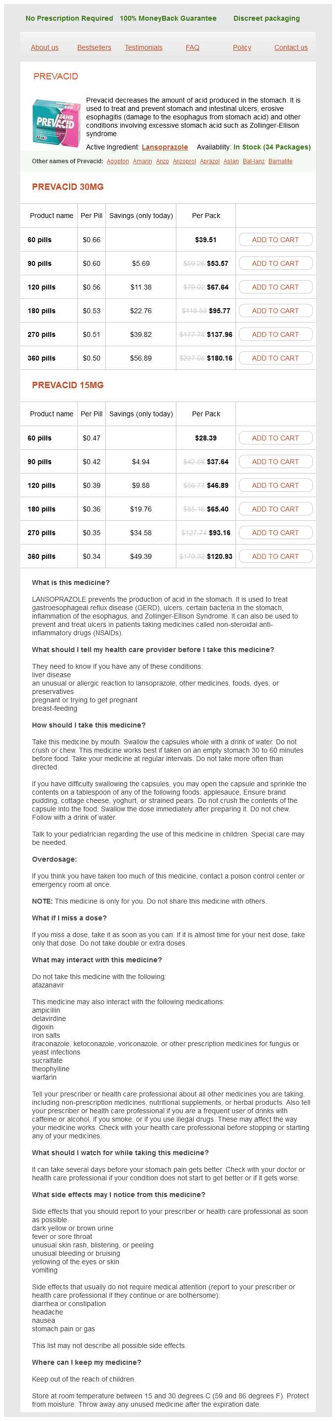
Lansoprazole, generally recognized by its model name Prevacid, is a proton pump inhibitor (PPI) medicine used to lower the amount of acid produced within the abdomen. It is primarily prescribed to deal with and prevent abdomen and intestinal ulcers, in addition to to alleviate symptoms of situations involving excessive stomach acid corresponding to erosive esophagitis and Zollinger-Ellison syndrome.
Prevacid can also be used to deal with a uncommon condition called Zollinger-Ellison syndrome, during which tumors in the pancreas and small gut trigger the abdomen to supply extreme quantities of acid. This can result in severe stomach ulcers and different digestive issues. Prevacid is prescribed to scale back the quantity of acid within the stomach, providing reduction from symptoms and serving to to forestall complications from the situation.
The fossa contains no important structures and can, therefore, be incised without fear when infected gastritis symptoms reflux purchase lansoprazole 15 mg on line. The female genital organs the vulva the vulva (or pudendum) is the term applied to the female external genitalia. The labia majora are the prominent hair-bearing folds extending back from the mons pubis to meet posteriorly in the midline of the perineum. The labia minora lie between the labia majora as lips of soft skin which meet posteriorly in a sharp fold, the fourchette. Anteriorly, they split to enclose the clitoris, forming an anterior prepuce and posterior frenulum. The vestibule is the area enclosed by the labia minora and contains the urethral orifice (which lies immediately behind the clitoris) and the vaginal orifice. The vaginal orifice is guarded in the virgin by a thin mucosal fold, the hymen, which is perforated to allow the egress of the menses, and may have an annular, semilunar, septate or cribriform appearance. Rarely, it is imperforate and menstrual blood distends the vagina (haematocolpos). At first coitus the hymen tears, usually posteriorly or posterolaterally, and after childbirth nothing is left of it but a few tags termed carunculae myrtiformes. Anteriorly, each gland is overlapped by the bulb of the vestibule a mass of cavernous erectile tissue equivalent to the bulbus spongiosum of the male. This tissue passes forwards, under cover of bulbospongiosus, around the sides of the vagina to the roots of the clitoris. This starts at the fourchette and extends posterolaterally on the right side for 1. The skin, vaginal epithelium, subcutaneous fat, perineal body and superficial transverse perineal muscle are incised. The cervix projects into the anterior part of the vault of the vagina so that the continuous gutter surrounding the cervix is shallow anteriorly (where the vaginal wall is 3 in (7. This continuous gutter is, for convenience of description, divided into the anterior, posterior and lateral fornices. The female genital organs 149 Relations · Anteriorly the base of the bladder and the urethra (which is embedded in the anterior vaginal wall). To make matters worse, this is the only part of the vagina which is covered by peritoneum; the peritoneal cavity is thus entered and peritonitis follows. Blood supply Arterial supply is from the internal iliac artery via its vaginal, uterine, internal pudendal and middle rectal branches. Structure A stratified squamous epithelium lines the vagina and the vaginal cervix; it contains no glands and is lubricated partly by cervical mucus and partly by desquamated vaginal epithelial cells. In nulliparous women the vaginal wall is rugose, but it becomes smoother after childbirth. The rugae of the anterior wall are situated transversely; this allows for filling of the bladder and for intercourse. This allows for sideways stretching to accommodate a rectum distended with stool and for the passage of the fetal head. Beneath the epithelial coat is a thin connective tissue layer separating it from the muscular wall, which is made up of a criss-cross arrangement of involuntary muscle fibres. This muscle layer is ensheathed in a fascial capsule that blends with adjacent pelvic connective tissues, so that the vagina is firmly supported in place. The Fallopian (uterine) tubes enter into each superolateral angle (the cornu), above which lies the fundus. The body of the uterus narrows to a waist termed the isthmus, continuing into the cervix which is embraced about its middle by the vagina; this attachment delimits a supravaginal and vaginal part of the cervix. The anatomical internal os marks its junction with the uterine body but its mucosa is histologically similar to the endometrium. The isthmus is that part of the uterus which becomes the lower segment in pregnancy. The cavity of the uterine body is triangular in coronal section, but in the sagittal plane it is no more than a slit. This cavity communicates via the internal os with the cervical canal, which, in turn, opens into the vagina by the external os. The nulliparous external os is circular, but after childbirth it becomes a transverse slit with an anterior and a posterior lip. The non-pregnant cervix has the firm consistency of the nose; the pregnant cervix has the soft consistency of the lips. In fetal life the cervix is considerably larger than the body; in childhood (the infantile uterus) the cervix is still twice the size of the body but, during puberty, the uterus enlarges to its adult size and proportions by relative overgrowth of the body. The adult uterus is bent forwards on itself at about the female genital organs 151 the level of the internal os to form an angle of 170°; this is termed anteflexion of the uterus. Moreover, the axis of the cervix forms an angle of 90° with the axis of the vagina anteversion of the uterus. In retroversion of the uterus, the axis of the cervix is directed upwards and backwards. Normally, on vaginal examination the lowermost part of the cervix to be felt is its anterior lip; in retroversion, either the os or the posterior lip becomes the presenting part. In retroflexion the axis of the body of the uterus passes upwards and backwards in relation to the axis of the cervix. They may be mobile and symptomless as a result of distension of the bladder or purely as a development anomaly.
Animals lacking the adenosine A2a receptor are hypoalgesic to heat stimuli (Ledent et al 1997) gastritis diet during pregnancy lansoprazole 15 mg purchase mastercard. Multiple purinergic (P2) receptors have been suggested to be involved in pain signaling and modulation. Local intradermal injection of agents activating P2X receptors results in dose-related pain behavior in rodents that is mediated by capsaicin-sensitive neurons (Bland-Ward and Humphrey 1997) and enhanced pain behavior in response to formalin (Sawynok and Reid, 1997). The proportion of C-fiber nociceptors responding and the magnitude of their response are increased by P2X agonists in inflamed skin. Serotonin causes pain when applied to a human blister base (Richardson and Engel 1986) and can activate nociceptors (Lang et al 1990). Serotonin can also potentiate the pain induced by bradykinin and enhance the response of nociceptors to bradykinin. The role of histamine in pain sensation is less clear since application of exogenous histamine to the skin produces itch and not pain sensations (Simone et al 1991a). Recently, it has been suggested that peripheral adenosine receptors may also be involved in the modulation of inflammatory pain. These studies provide evidence for a role of cytokines in inflammation-associated hyperalgesia. Several lines of evidence indicate a role of peripheral mGluRs in nociception and inflammatory pain. Peripheral application of glutamate activates nociceptors, and peripheral administration of ligands binding to glutamate receptors induces pain behavior in animals. Involvement of peripheral iGluR, mGluR1, and mGluR5 in formalin-induced pain behavior and glutamate-induced thermal hyperalgesia has been demonstrated (Davidson et al 1997). Intraplantar, but not intrathecal or intracerebroventricular administration of an mGluR5 antagonist reduced inflammatory hyperalgesia. Endogenous sources of glutamate in the periphery include plasma, macrophages, epithelial and dendritic cells in the epidermis and dermis, and Schwann cells. In addition, peripheral processes of the primary afferents contain glutamate, and nociceptor stimulation can cause peripheral release of glutamate from the terminals of these afferents. Endothelin Receptors Endothelins are vasoactive peptides that are widely distributed in somatic and visceral tissue (for reviews see Hans et al 2009, Khodorova et al 2009). Endothelins have been implicated in the pain and hyperalgesia associated with inflammation, skin incision, cancer, and sickle cell crisis. Other Receptors A number of other receptor systems have been reported to play a role in the peripheral modulation of nociceptor responsiveness. Drugs acting at the periphery and inhibiting the generation and signaling of nociceptive input toward the spinal cord and the brain may prevent central plastic changes such as wind-up and central sensitization. Opioid receptors have been demonstrated on the peripheral terminals of afferent fibers, and axonal transport of these receptors is enhanced during inflammation. Peripheral analgesia by opioids appears to be part of a physiological antinociceptive system since increased amounts of endogenous opioids have been found in inflamed tissues. Inflammatory cells such as macrophages, monocytes, and lymphocytes contain opioid peptides. An alternative mechanism for activation of endogenous opioid analgesia at the site of tissue injury has been described (see Khodorova et al 2009 for review). Cholinergic Receptors Non-neuronally released acetylcholine, acting on peripheral cholinergic receptors, may have a modulatory role on nociception. Nicotine has a weak excitatory effect on C-fiber nociceptors and induces mild sensitization to heat, but no alterations in mechanical responsiveness. In contrast, muscarine desensitizes C nociceptors to mechanical and heat stimuli (Bernardini et al 2001). Thus, nicotinic and muscarinic receptors may have opposing effects on cutaneous nociceptors. Studies in mice with targeted deletions of the M2 receptor gene suggest that M2 receptors on cutaneous nerve endings depress the responsiveness of nociceptive fibers to noxious stimuli (see Wess et al 2003 for review). M2, M3, and M4 muscarinic receptor subtypes may be involved in the modulation of nociceptive transduction. However, peripheral 2-adrenoceptors may also be involved in modulation of nociceptor activity. Studies using selective 2-subtype knockout mice have shown that the 2Aadrenergic receptors are primarily involved in the analgesic effect of 2-adrenoceptor agonists (Stone et al 1997). Cannabinoids Cannabinoids have recently emerged as a potential therapy for chronic pain. They are distributed at many key sites in the pain-signaling pathway, including the peripheral and central terminals of primary afferent fibers, spinal dorsal horn neurons, and the brain stem and brain. Although some of these agents may directly activate nociceptors, most of the inflammatory mediators lead to changes in the sensory neuron rather than directly activating it. Such changes in sensory neurons include early post-translational alterations in the peripheral terminals of nociceptors (peripheral sensitization) and a delayed transcription-dependent alteration (see Woolf and Costigan 1999, Kidd and Urban 2001). Considerable attention has been focused on the signal transduction mechanisms of primary afferent neurons and their alteration by inflammation. Two principal signaling pathways have been postulated to mediate inflammation-induced hyperalgesia. As with other types of tissue injury, secondary hyperalgesia after incision injury is present only to mechanical, not thermal, stimuli (Pogatzki et al 2000). The incision-induced primary and secondary hyperalgesia results from characteristic peripheral, spinal, and supraspinal mechanisms (Zahn and Brennan 1999, Pogatzki et al 2002).
Lansoprazole Dosage and Price
Prevacid 30mg
- 60 pills - $39.51
- 90 pills - $53.57
- 120 pills - $67.64
- 180 pills - $95.77
- 270 pills - $137.96
- 360 pills - $180.16
Prevacid 15mg
- 60 pills - $28.39
- 90 pills - $37.64
- 120 pills - $46.89
- 180 pills - $65.40
- 270 pills - $93.16
- 360 pills - $120.93
Like heat, repeated presentations of a mechanical stimulus lead to pronounced fatigue gastritis zyrtec lansoprazole 15 mg buy visa. Much has been learned about the features of a mechanical stimulus that determine the response of nociceptors to mechanical stimuli. The discharge of nociceptors increases with increased force and pressure, but these functions vary depending on probe size: the smaller the probe, the greater the response (Garell et al 1996). For cylindrical probes of different diameter, the discharges are comparable if the intensity of the stimulus is calculated according to force per length of the perimeter of the cylindrical probe. This suggests that the stress/strain maximum that occurs at the edge of the cylindrical stimulus is the critical parameter for excitation of nociceptor terminals. For a given probe size, the response of A-fiber nociceptors increases monotonically with force, whereas the response of C-fiber nociceptors becomes saturated at higher force levels. Mechanical stimuli were presented to the receptive field of A-fiber and C-fiber nociceptors at different interstimulus intervals (with 10 minutes between stimulus pairs). The A-fiber response (triangles) recovered within 60 seconds, whereas the C-fiber response (circles) took more than 150 seconds to recover. To normalize the data, the response to the test stimulus was divided by the response to the immediately preceding conditioning stimulus. However, the transducer elements that account for mechanosensitivity are probably different from those responsible for heat. For example, the heat response of nociceptors is readily sensitized by a heat injury, whereas the mechanical response is not (see below). A-fiber nociceptors appear to be responsible for the sharp pain reported in response to punctate mechanical stimuli: (1) the reaction time to perception of pain is short, (2) the stimulusresponse function of A-fiber nociceptors. Pretreatment of the skin with capsaicin abolishes heat pain sensitivity but does not greatly affect mechanical pain (Magerl et al 2001). Comparison of responses of nociceptors to mechanical stimuli in the monkey with pain ratings in human subjects. These data provide evidence that A-fiber nociceptors signal the pain reported from sharp probes. A, Average responses of A-fiber nociceptors (triangles) and C-fiber nociceptors (circles) to controlled-force stimuli. The A fibers exhibited a monotonically increasing response, whereas the response of the C fibers reached a plateau at the higher force levels (0. B, Average pain ratings in response to controlled-force stimuli (open circle) increased monotonically in a manner comparable to that observed for the A-fiber nociceptors. Selective block of A-fiber function led to a significant decrease in pain ratings (filled circles). Klement and Arndt (1992) demonstrated that cold pain could be evoked by cold stimuli applied within the veins of human subjects. A local anesthetic applied within the vein, but not in the overlying skin, abolished cold pain sensibility. It is therefore possible that cold pain is served, at least in part, by vascular receptors. Just as the sensation of warmth is served by a specific set of primary afferents (predominantly C fibers), the sense of cooling is served by a specific set of primary afferents. They exhibit ongoing activity at room temperature, and their response increases markedly with gentle cooling. Although the majority of nociceptors have some response to ice stimuli applied to the skin, Simone and Kajander (1997) showed that all A-fiber nociceptors respond to cold stimuli below 0°C. C-fiber nociceptors may play a role in signaling cold pain sensation as well (LaMotte and Thalhammer 1982). This channel is found in a subset of nociceptive sensory neurons that are responsive to intense heat and capsaicin. There is, in fact, a dynamic plasticity that relates stimulus intensity and sensation. Hyperalgesia is defined as a leftward shift of the stimulus response function that relates the magnitude of pain to stimulus intensity. It is evident that the threshold for pain is lowered and pain in response to suprathreshold stimuli is enhanced. Hyperalgesia is a consistent feature of somatic and visceral tissue injury and inflammation. Pharyngitis is associated with hyperalgesia in pharyngeal tissues such that merely swallowing induces pain. Micturition in the presence of a urinary tract infection is painful, again reflecting the presence of hyperalgesia. The peripheral neural mechanisms of hyperalgesia have been studied in various tissues, including the joints, cornea, testicle, gastrointestinal tract, and bladder. Much of the theoretical work on hyperalgesia, however, has evolved from studies of the skin, and it is this work that will receive attention here. Hyperalgesia occurs not only at the site of injury but also in the surrounding uninjured area. Hyperalgesia at the site of injury is termed primary hyperalgesia, whereas hyperalgesia in the uninjured skin surrounding the injury is termed secondary hyperalgesia (Lewis 1935). As we will see, the neural mechanisms for primary and secondary hyperalgesia differ. In discussing hyperalgesia, it is useful to consider the following variables: (1) energy form of the injury, (2) type of tissue involved, (3) energy form of the test stimulus, and (4) location of the testing relative to the area injured. For example, it will be shown that nociceptors will become sensitized to mechanical stimuli (the energy form of the test stimulus), but only after certain forms of injury. An experimental design frequently used for study of the neural mechanisms of hyperalgesia is to characterize the response properties of a given fiber, then apply a manipulation that under usual circumstances would produce hyperalgesia, and finally assess whether this manipulation has altered the response properties of the fiber in question.

ADDII BIOTECH is proud to announce that the company's manufacturing facility located at Baddii, Himachal Pradesh has recently been approved by Food & Drug Authority of Ghana.
Our Manufacturing capability includes a wide range of therapeutic products covering almost every segment.
ADDII BIOTECH given our strong emphasis on product quality and services.