
Lithium
General Information about Lithium
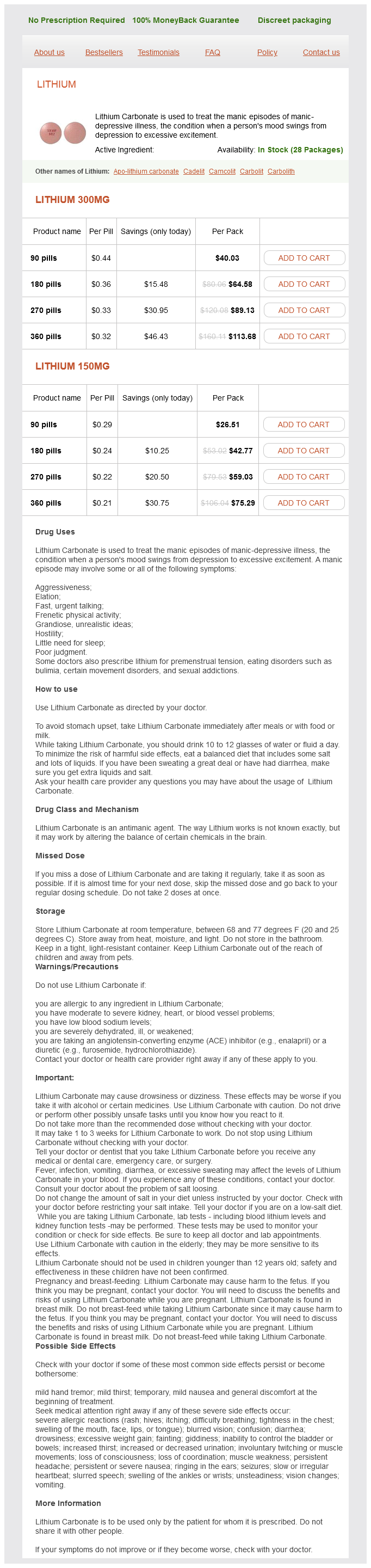
The most commonly used type of lithium for treating bipolar disorder is lithium carbonate. It is available in several forms, including tablets, capsules, and extended-release tablets. The dosage is carefully monitored and tailor-made to every particular person to achieve the best outcomes.
While lithium is very effective in treating bipolar disorder, it does come with some side effects. These can include frequent urination, dry mouth, nausea, and tremors. Therefore, it is essential for people taking lithium to obtain common check-ups and blood checks to ensure that the dosage is appropriate and that their levels are inside a safe range.
One of the most well-known makes use of of lithium is in the therapy of manic-depressive or bipolar disorder. This mental illness is characterised by extreme temper swings, with the person experiencing intervals of melancholy adopted by durations of high power and pleasure. While the exact cause of bipolar dysfunction continues to be unknown, scientists consider that imbalances in mind chemical substances, similar to serotonin and dopamine, play a key position. This is where lithium is available in.
Furthermore, lithium has additionally been used in the remedy of physical ailments similar to gout, certain types of complications, and fibromyalgia. It has anti-inflammatory properties and has been shown to improve brain operate and reminiscence in older adults.
So, how does lithium work? It is believed that lithium helps to regulate the levels of neurotransmitters, corresponding to serotonin and dopamine, within the mind. This might help to stabilize the acute mood swings and scale back symptoms of each melancholy and mania. It also has a calming impact on the mind, which can help to reduce agitation, irritability, and impulsivity.
In addition to treating bipolar dysfunction, lithium has also been found to be useful in treating other mental well being conditions similar to schizophrenia and major depressive disorder. It has also proven promise in preventing suicide in people with bipolar dysfunction.
Lithium, a soft and silvery metallic, has been used for tons of of years in its varied forms for a broad array of functions. From powering batteries to treating psychological illness, lithium has proven to be a flexible factor with many functions.
In conclusion, lithium is a crucial factor with many uses and applications. From treating psychological illness to powering our devices, lithium has proven to be a flexible and priceless resource. However, its effectiveness in treating bipolar disorder cannot be overlooked, as it has considerably improved the standard of life for countless people dwelling with this challenging situation. With more research and developments, the potential of lithium within the medical and technological fields is limitless.
Aside from its psychiatric makes use of, lithium can also be broadly used within the manufacturing of batteries. In truth, it's the lightest steel and has the best electrochemical potential of all the weather. This makes it ideal to be used in rechargeable batteries, such as those found in cell phones, laptops, and electrical automobiles.
In the 19th century, French psychiatrist Jean Pierre Falret first noticed the effectiveness of lithium in treating signs of manic-depressive illness. However, it wasn't till the Forties that it was formally recognized as a remedy choice. Today, lithium is considered to be the gold commonplace in the remedy of bipolar disorder.
Other surgical procedures including epikeratophakia symptoms 24 hours before death buy generic lithium from india, hexagonal keratotomy, automated lamellar keratoplasty, and thermal keratoplasty have all been abandoned due to the significant complications related to each procedure. There was one case of malignant glaucoma which occurred 2 years after surgery in an eye that had previously had a Yag iridectomy. Although one eye experienced rupture of the posterior capsule, no eye lost any vision permanently. This technique is often considered an option when treating higher degrees of ametropia. The advancement of preoperative biometry as well as intraocular lens power formulas have resulted in excellent postoperative refractive results. For hyperopic patients younger than 40 years, phakic intraocular lens implantation may be a better option in order to preserve accommodation if their anterior chamber is of adequate depth. Seiler T: Clear lens extraction in the 19th century an early demonstration of premature dissemination. Mimouni F, Collin J, Koffi V, Bonnet P: Damage to the corneal endothelium from anterior chamber intraocular lenses in phakic myopic eyes. Kohnen S, Brauweiler P: First results of cataract surgery and implantation of negative power intraocular lenses in highly myopic eyes. Colin J, Robinet A: Clear lensectomy and implantation of a low-power posterior chamber intraocular lens for correction of high myopia: a four-year follow-up. Nishi O, Nishi K, Sakanishi K: Inhibition of migrating lens epithelial cells at the capsular bend created by the rectangular optic edge of a posterior chamber intraocular lens. Buehl W, Findl O, Menapace R, et al: Effect of an acrylic intraocular lens with a sharp posterior optic edge on posterior capsule opacification. Pucci V, Morselli S, Romanelli F, et al: Clear lens phacoemulsification for the correction of high myopia. Colin J, Robinet A, Cochener B: Retinal detachment after clear lens extraction for high myopia: seven-year follow-up. Colin J, Robinet A: Clear lensectomy and implantation of low-power posterior chamber intraocular lens for the correction of high myopia. Mastropasqua L, Carpineto P, Ciancaglini M, et al: Treatment of retinal tears and lattice degenerations in fellow eyes in high risk patients suffering retinal detachment: a prospective study. Preetha R, Goel P, Patel N, et al: Clear lens extraction with intraocular lens implantation for hyperopia. Only when it was considered as a refractive procedure, were methods developed to improve intermediate and near vision. After 45 years of age, crystalline lens surgery is considered to be one of the most popular surgeries between the increasingly educated patients demanding improved functional vision in other words, improved far and near vision. This article considers the currently available models of these lenses that are in varying stages of development and capable of providing near vision. The recent models of the lenses included in this chapter are divided into two main groups: 1. Fixation within the capsular bag in ensured by the presence of small, Tshaped polyimide haptics at the end of the plates. When the ciliary muscle constricts, it redistributes its mass like any other muscle and encroaches on the vitreous cavity space, increasing the vitreous cavity pressure, moving the optic forward. An increase in vitreous cavity pressure thus moves the optic forward; 1 mm of movement is equivalent to almost a 2 D power change. In the nonaccommodative phase, the tension of the capsular bag and zonules keeps the two optics in close proximity, whereas the spring devices are collapsed and exhibit potential energy. With accommodative effort, the zonules relax, the capsular bag expands, and the springs express kinetic energy. This change allows the optics to separate as the anterior plus lens moves forward, thus producing a higher optical power that yields accommodation. The mechanism of action of this lens is based on a lens complex formed by two optics linked by a spring system. The principle of action of the lens is focus shift during the anterior movement of the optic secondary to ciliary muscle contraction. In addition, the flexibility of the capsular bag remains an important aspect of performance for this lens design. Kellen Tetraflex Accommodating Lens Design: the lens haptic was designed to take advantage of how the crystalline lens moves during accommodation according to the Helmholtz theory. This theory states that contraction of the ciliary muscles relaxes the lens zonules, thereby allowing the lens to move forward during accommodation. It is not designed on a hinge principle, but rather on a haptic configuration to allow the lens to move with the entire capsular bag. Accommodative and Pseudoaccommodative Intraocular Lenses under tension) to a maximum of 2. During accommodation, the contracting ciliary body will result in zonular relaxation, releasing the tension on the capsular bag and allowing release of the spring and an increase in the interoptical distance, which leads to anterior optic forward shift. The posterior lens is designed with a significant large area so as to reduce the tendency toward posterior axial excursion and maintain stability and centration within the capsular bag at all times. The optical power of the anterior element of the system is +30 to +35 D and it is the posterior element that has a negative variable power to bring the eye to emmetropia according to patient characteristics. Based upon the application of applied microfluid dynamics models, this lens has a peripheral fluid reservoir. Upon accommodative stimulation, an actuator triggers microscopic pumps to move fluid from the periphery to the center of the lens, thereby increasing its anterior/posterior dimension and, hence, its optical power. The haptic design has more than one function: the haptics fix the lens system all the time in the capsular bag and it then provides a spring-like resistance separating the two optics of ~2. The lens when implanted in the capsular bag occupies the entire capsule and uses the contraction and relaxation of the ciliary muscle against the spring force of the haptic to emulate accommodation of the natural human lens. Its single optic is located in the anterior-most part of the lens and this will be situate the optic in an anteriorly biased location within the capsular bag.
Reprinted from Piatigorsky J: Gene expression and genetic engineering in the lens symptoms 4 months pregnant proven lithium 150 mg. Invest Ophthalmol Vis Sci 1987; 28:9 Lens Proteins and Their Molecular Biology key motifs, is present in all b-crystallins. The first and second motifs are encoded in the second exon and the third and fourth motifs are encoded in the third exon in all the gcrystallins. However, the first three codons of the g-crystallin polypeptides are situated in a small 5, exon (exon 1) in a fashion similar to that of the amino terminal arms of the b-crystallins. With the exception of gS-crystallin, which is located on chromosome 3pter in humans, the g-crystallin genes are found in a cluster on chromosome 2q33 (Table 105. In humans, gE-crystallin and gF-crystallin are pseudogenes with early termination codons and inactive or absent promoters, while the ygG-crystallin pseudogene maintains only a fragment of the g-crystallin gene structure. The gC-, gD-, and gS-crystallins comprise the bulk of the g-crystallins in the human lens with gA- and gB-crystallins being expressed at lower levels. The phylogenetic and developmental distribution of the g-crystallins differs from that of the b-crystallins. All members of the b-crystallins are well represented in all vertebrate lenses (although their proportional representation and spatial distribution within the lens vary with species), while the g-crystallins are poorly represented in birds and reptiles. Indeed, it used to be thought that g-crystallins were entirely absent from lenses of birds and reptiles, but the finding that some reptiles contain g-crystallins98 and that the gS polypeptide (formerly called bs) present in birds and reptiles is more g-like than b-like on the basis of its gene structure (discussed later),99 has modified the extreme view that g-crystallins are excluded from the bird and reptile lineages. In general, the g-crystallins other than gS are synthesized primarily in the early stages of development so that the content of g-crystallin is greater in the lens nucleus than in the younger cortical regions of the lens. Similarly, the g-crystallin content is higher in the more rigid lenses of species such as rodents and fish, which do not accommodate by changing lens shape. This suggests that the g-crystallins have been adapted for regions with very high-density molecular packing rather than more hydrated regions of lower protein concentration. Association of b-crystallins into dimers and higher order aggregates appears to be a complex process. The surface interactions between b-crystallin monomers associating to form a dimer are similar to those of two g-crystallin domains associating intramolecularly. The terminal arms do not appear to be required for association of mouse bA3/A1-crystallin or bovine bB2-crystallin to form homodimers. Reprinted from Bax B, Lapatto R, Nalini V, et al: X-ray analysis of beta B2-crystallin and evolution of oligomeric lens proteins. In bB2-crystallin crystals, although not in bB1113 this connecting peptide maintains an extended conformation, requiring intermolecular association of two b-crystallins into dimers. While replacing the connecting peptide in gB-crystallin with the bB2 linker does not alter the monomeric g-crystallin structure,114 placing a gB-crystallin connecting peptide in b-crystallins results in monomeric bovine bB2 but not mouse bA1/A3-crystallins. Rather, association of the b-crystallin polypeptides appears to be entropically driven, probably by monomers imposing increased order on the surrounding hydration shell. For example, although it is located on a different chromosome than the other g-crystallins, the gS gene has a single exon for each domain (comprising two motifs) like the other g-crystallin genes rather than separate exons for each structural motif, like the b-crystallin genes. This is true as well for gN-crystallin, which has an amino terminal arm as gS-crystallin and the b-crystallins. Although weakly linked to the b-crystallins, its amino acid sequence groups separately from the b- and other g-crystallins. Of particular interest, the gN-crystallin gene encodes the first protein domain comprising two Greek key motifs in one exon like the other g-crystallin genes, but encodes the next two Greek key motifs comprising the second domain in separate exons. It is present in vertebrates, but there is no evidence for its expression in primates, where it might be a pseudogene. While many of the bg-crystallins appear to be lens-specific, several members of both families are expressed in retina and a variety of other tissues,124128 the functional significance of nonlens bg-crystallins is not known yet. However, homologs from the wider bg-crystallin superfamily suggest roles related to growth and physiological stress. A second nonlens member of this family is spherulin 3A of the slime mold Physarum polycephalum,132 a microbial protein required for encystment and dormancy. Spherulin 3A has a sequence which is consistent with a tertiary structure resembling a single domain of the bg-crystallins. Once more, the order of fusion of motif structures into the domain is reversed between spherulin 3A and the bg-crystallins. While these two distant members of the bg-crystallin superfamily are microbial coat proteins, it is possible that they play a more general role as protective stressinduced proteins. In this regard both proteins are produced in response to osmotic stress (in contrast to sporulation in many organisms), providing a functional connection with the a-crystallin family which is homologous to the small heat shock proteins. The sequence similarity between the Ci-bg-crystallin and vertebrate bg-crystallins is weak, but their crystal structures are strikingly related, suggesting that Ci-bg-crystallin is a direct precursor to the vertebrate crystallins. This is consistent with the fact that larval urochordates gave rise to the vertebrate lineage. Ci-bg-crystallin is expressed in the palps (substrateadhesive structures) and the otolith (a sensory, opsin-containing structure in brain) of the urochordate larva and clearly does not perform a refractive role as do its homologs in the vertebrate lens. Most of these crystallins are related or identical to functional enzymes which occur at low concentrations in nonlens tissues. There are numerous enzyme-crystallins found in different species of all vertebrate and invertebrate classes (Table 105. Some are products of single copy genes while others have sustained one or more gene duplications. Diagram showing different ways by which recruitment of enzymes as lens crystallins might occur. A housekeeping gene expressed in many tissues (top) may be modified by a number of mechanisms, increasing its expression in the lens while maintaining low levels of expression in other tissues (bottom). Reprinted from Piatigorsky J: Lens crystallins and their genes: diversity and tissue-specific expression. Cephalopods express W-crystallin, which was derived from an ancestral aldehyde dehydrogenase gene that lost enzymatic activity as it became specialized for lens expression,146 and S-crystallins, which are inactive glutathione-S-transferases (except for one; discussed later).
Lithium Dosage and Price
Lithium 300mg
- 90 pills - $40.03
- 180 pills - $64.58
- 270 pills - $89.13
- 360 pills - $113.68
Lithium 150mg
- 90 pills - $26.51
- 180 pills - $42.77
- 270 pills - $59.03
- 360 pills - $75.29
Cilioretinal arteries arise from these short posterior ciliary arteries and are present in 30% of eyes and 50% of individuals symptoms 10 dpo 300 mg lithium purchase visa. The laminar and prelaminar optic nerve is supplied by branches of the posterior ciliary arteries. The deeper optic nerve in supplied by intraneuronal branches from the central retinal artery and recurrent pial branches of the central retinal and ophthalmic artery. Within the retina, larger branches of the central artery course through the nerve fiber and ganglion cell layers. As the arterial circulation divides and lumen size narrows smaller arterioles and capillaries extend through these layers into the inner nuclear layer. While retinal circulation is generally end arterial in nature, two anastomoses exist in the region of the optic disk between branches of the central retinal artery and short posterior ciliary arteries. There intraneuronal branches of the central retinal artery communicate with recurrent pial branches. The decrease or absence of perfusion caused by central retinal artery obstruction leads to decreased delivery of glucose and more importantly oxygen6 to the tissues of the retina. Ischemia leads to edema as neuron cellular membranes burst4,7 producing the associated funduscopic whitening of the retina caused by opacification of the ganglion cell layer. The opacification is most prominent in the macular region where the multiple layers of ganglion cells reside. Retinal opacification generally decreases toward the periphery as the ganglion cell population diminishes, and is also absent from the avascular zone of the foveola where ganglion cells are absent. Animal studies have demonstrated swelling of the organelles that precede axonal and dendritic rupture with decreased neuronal cell count. Common systemic risks factors for retinal arterial occlusive disease include hypertension, diabetes, lipid disorders, and cardiac and systemic atherosclerotic disease. It is difficult to determine the precise incidence and prevalence of arterial occlusive events in the retina. Other mechanisms such as radiation and elevated intraocular pressure occur much less commonly. It is important to note that more than one mechanism may occur at the same time in a patient. An embolism to the retinal arterial circulation can originate as a venous thrombosis reaching the arterial circulation via a right to left cardiac shunt. Patients aged 50 and greater are likely to have occlusions associated with embolic disease while younger patients are more likely to have occlusions secondary to thrombosis. Atherosclerosis of the carotid system is the most common source of retinal emboli with 80% being associated with carotid artery disease. Although degree of obstruction is important, the presence of an ulcerative plaque, even with minimal stenosis, can be more likely to lead to emboli than an extensively occluded carotid artery. The heart and major vessels are another important source of emboli that must be considered when confronted with a patient with embolic retinal arterial occlusions. Valvular abnormalities,1921 tumors, like atrial myxomas,22,23 and thrombus formation in the left atrium secondary to conditions like atrial fibrillation, can lead to emboli formation and vascular occlusion. Additionally, right to left shunts, like patent foramen ovale, can allow emboli from venous thrombotic disease to reach the arterial circulation. Note that there is normal reflectivity of the retinal pigment epithelium at the foveola. Retinal Arterial Occlusions disease originating from the heart is echocardiography. In cases where there is a high suspicion of disease that is not detected by transthoracic echocardiography, transesophageal echocardiography can be helpful in identifying a source of emboli. Emboli/Exogenous Source Exogenous emboli can reach the retinal circulation leading to arterial occlusive events. Examples include talc emboli associated with intravenous drug abuse30 and emboli from injections of steroids in the nasal or periorbital area,31,32 or related to blood products like platelet emboli during transfusion. Fragments of catheter tips and artifical heart valves may also cause arterial occlusion. Retinal Emboli/Types Although embolic obstruction is presumably the most common cause of retinal arterial occlusions, visible emboli detected ophthalmoscopically are only evident in 2030% of patients with retinal arterial occlusive disease. Cholesterol emboli are small, glistening yellow in color, and typically do not occlude the vessel distal to the emboli. Platelet fibrin emboli may appear gray-white, are larger and longer than cholesterol emboli, and may be seen to pass through the retinal vessels. Calcific emboli are larger, gray-white in color, and associated with more severe local obstructions. Arruga felt that calcific emboli were more likely to arise from cardiac valvular disease. While venous thrombosis occurs most commonly, arterial thrombotic disease of the retina also does occur. Structural abnormalities in a vessel like prepapillary arterial loop can contribute to retinal arterial occlusion by thrombosis. Thrombophilia is the term used to describe conditions that are genetically determined and increase the likelihood of vascular thrombosis (Table 131. These abnormalities typically lead to venous thrombosis, but when they interact with other local or systemic hypercoagulable factors like trauma, malignancy, pregnancy, oral contraceptive use, autoimmune disease, and cigarette smoking, they may lead to arterial thrombosis. These hereditary thrombophilic states should be considered in patients with retinal arterial occlusions, particularly with a positive family history of thrombotic events occurring at a young age. While entire textbooks have been devoted to discussing these thrombophilic conditions, a brief explanation of some of the more common of these conditions that have been associated with retinal arterial occlusive events will be useful.

ADDII BIOTECH is proud to announce that the company's manufacturing facility located at Baddii, Himachal Pradesh has recently been approved by Food & Drug Authority of Ghana.
Our Manufacturing capability includes a wide range of therapeutic products covering almost every segment.
ADDII BIOTECH given our strong emphasis on product quality and services.