
Micronase
General Information about Micronase
Micronase works by stimulating the beta cells of the pancreas to supply extra insulin. It also helps the body's cells to use insulin more successfully. By doing so, it helps to decrease blood sugar levels and hold them within a traditional range. This can reduce the risk of growing severe issues of diabetes, such as heart illness, nerve harm, and kidney injury.
It is essential to tell a health care provider about all present drugs and medical circumstances earlier than beginning remedy with Micronase. This treatment might interact with certain medications, corresponding to blood thinners, and is most likely not appropriate for individuals with specific medical conditions, such as liver or kidney illness. It can additionally be not beneficial to be used throughout pregnancy or while breastfeeding.
Type 2 diabetes is a continual situation by which the physique is unable to properly regulate the levels of glucose (sugar) within the blood. This occurs when the physique turns into resistant to the consequences of insulin or doesn't produce sufficient insulin to meet the physique's wants. Insulin is a hormone that is produced by the pancreas and performs a crucial role in regulating blood sugar ranges. When the body is unable to supply or use insulin effectively, it may possibly result in excessive ranges of glucose within the blood, which can cause critical health complications.
The treatment is on the market in tablet type and is normally taken a couple of times a day, relying on the person's wants. It is important to take it exactly as prescribed by a healthcare skilled. The beneficial beginning dose is normally 2.5 mg per day, which can be elevated gradually relying on the response of the individual. Micronase must be taken with meals to forestall low blood sugar levels (hypoglycemia). It is important to observe blood sugar ranges frequently whereas taking this medicine to make sure it's working successfully and not inflicting any adverse results.
In conclusion, Micronase is an efficient medication for the administration of type 2 diabetes. It helps to manage blood sugar ranges and cut back the chance of issues associated with this situation. However, it is not a remedy for diabetes, and life-style modifications, similar to healthy eating and common exercise, are also crucial in its management. It is crucial to work intently with a healthcare professional to determine the most effective therapy plan for a person's particular needs. With proper use and monitoring, Micronase can enhance the quality of life for these residing with sort 2 diabetes.
Micronase is a protected and well-tolerated medicine for most people. However, like all drugs, it could cause some side effects. The mostly reported facet impact is low blood sugar (hypoglycemia), which can trigger symptoms such as dizziness, sweating, confusion, and faintness. To forestall this, it could be very important eat common meals and snacks and to verify blood sugar ranges frequently. Other possible side effects could include nausea, vomiting, abdominal ache, and headache. In rare cases, allergic reactions might happen, and immediate medical attention must be sought if any symptoms of an allergic response are skilled.
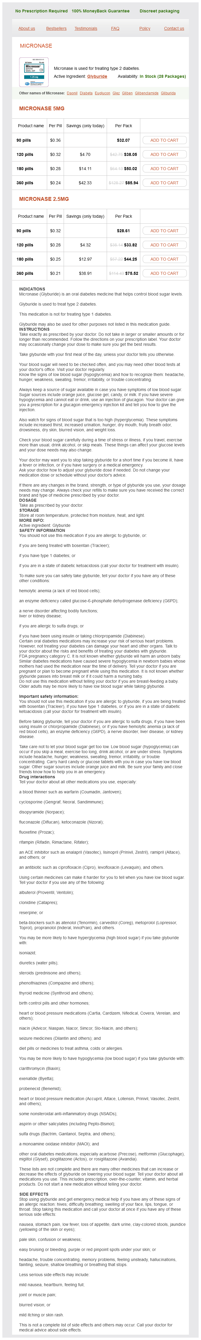
Micronase, additionally known by its generic name glyburide, is a broadly used medicine for the treatment of type 2 diabetes. It belongs to the group of medicines known as sulfonylureas, which work by rising the manufacturing of insulin in the pancreas and helping the body successfully use the insulin it produces. This treatment has been in use for over 50 years and is considered one of the efficient treatments for sort 2 diabetes.
Boffetta Anatomy the nasal cavities are separated in the midline by the nasal septum diabetes type 1 food chart 2.5 mg micronase purchase with mastercard. The floor is the hard palate, formed by the palatine processes of the maxillae and the horizontal portions of the palatine bones. The lateral nasal wall contains the maxillary and ethmoid ostia, plus three or four turbinates. These turbinates are delicate scroll-like projections of bone and vascular soft tissue that become smaller as they ascend in the nasal cavity. They attach to the lateral nasal wall anteriorly, and have a free edge posteriorly. The turbinates are covered with a thick mucous membrane and contain a dense, thick-walled venous plexus. The upper margins of the nasal fossa are bound laterally by the superior nasal turbinate and adjacent lateral nasal wall, and medially by the nasal septum. This mucosa contains bipolar olfactory nerve fibers that cross through the cribriform plate. The terminal axons of the olfactory nerves extend to the free surface of the epithelium, where they expand into knob-like protrusions bearing cilia (olfactory cilia). The nasal cavity and paranasal sinuses are lined by Schneiderian mucosa, consisting of pseudostratified columnar ciliated epithelium with interspersed goblet cells. The lamina propria within the paranasal sinuses, especially the maxillary antrum, is loose and well vascularized, with seromucinous glands, and can easily become polypoid as a result of edema. The goblet cell component of the mucosal surface and seromucinous glands is variable. In chronic sinusitis, goblet cell hyperplasia can result in a papillary mucosal lesion. The major portion of the nasal septum is formed by the perpendicular plate of the ethmoid bone posteriorly and the septal cartilage anteriorly. The septum is lined by relatively thin, ciliated respiratory mucosa, which may regularly undergo squamous metaplasia. The underlying thin lamina propria, although containing seromucinous glands, is tethered to the septal cartilage, restricting reactive polyp formation. The frontal sinus these paired sinuses reside between the internal and external cranial tables and drain either via a nasofrontal duct into the frontal recess or more directly into the anterior infundibulum, or less often, into the anterior ethmoid cells, which in turn will open into the infundibulum of the bulla ethmoidalis. Ethmoid complex this paired complex of sinuses contains 3-18 cells that are grouped as anterior, middle, or posterior, according to the location of their ostia. The left and right groups of ethmoid cells are connected in the midline by the cribriform plate (nasal roof) of the ethmoid bone. The cribriform plate is an important landmark in evaluation of sinonasal tumour stage - violation of the cribriform plate signifies direct extension of the tumour into the anterior cranial fossa. The crista galli is a distinctive pointed bony landmark that extends from the midline of the cribriform plate upward into the floor of the anterior cranial fossa. The perpendicular plate of the ethmoid bone extends downward from the cribriform plate to contribute to the nasal septum. The medial wall of each ethmoid labyrinth is formed by a thin lamella of bone from which arise the middle, superior, and supreme turbinates. The lateral ethmoid wall is formed by the thin lamina papyracea, which separates the ethmoid cells from the orbit. Tumour violation of the lamina papyracea may necessitate including the orbit and globe with the surgical resection. This area should be sampled in a maxillectomy specimen a) if the globe has not been removed (as the lamina papyracea represents the lateral orbital margin), or b) if orbital exenteration has been performed. The roof of the ethmoid complex is formed by a medial extension of the orbital plate of the frontal bone, which projects to articulate with the cribriform plate. Sphenoid sinus the average adult sphenoid sinus measures 20 mm high, 23 mm long, and 17 mm wide. The relationship of the posterior extension of the sphenoid in relation to the sella turcica is variable. The sphenoid sinus septum is usually in the midline, and anteriorly aligned with the nasal septum. With the exception of the sinus roof, the other sinus walls are of variable thickness depending on the degree of pneumatization. The sinus roof relates to the floor of the anterior cranial fossa, anteriorly; the optic chiasm and. The lateral sphenoid wall is related to the orbital apex, the optic canal, the optic nerve, and the cavernous sinus, containing the internal carotid artery. The sinus floor is the roof of the nasopharynx, and the anterior sinus wall is the back of the nasal fossa. Behind the orbital rims, each sinus roof/orbital floor slants obliquely upward so that the highest point of the sinus is in the posteromedial portion, lying directly beneath the orbital apex. The medial antral wall is the inferior lateral wall of the nasal cavity ("party wall"). The anterior sinus wall is the facial surface of the maxilla that is perforated by the infraorbital foramen below the orbital rim. The floor of the sinus is lowest near the second premolar and first molar teeth and usually lies 3-5 mm below the nasal floor. This pattern of drainage in the erect position is accomplished by intact ciliary action. The maxillary hiatus is a bony window leading to the interior of the maxillary sinus. The hiatus is normally partially covered by portions of four bones: the perpendicular plate of the palatine bone, posteriorly; the lacrimal bone, anterosuperiorly; the inferior turbinate, inferiorly, and above the turbinate attachment, the uncinate process of the ethmoid bone. The margin (black arrow) of the tumour in the maxillary sinus is separable from the obstructed secretions because of different densities.
Some attempts have been made to grade the spectrum of intraepithelial neoplastic changes (dysplasia/carcinoma-in situ blood sugar 70 after eating buy micronase in india, or nasopharyngeal intraepithelial neoplasia) in the nasopharynx, but reproducibility and difficulties in recognizing the lower grade lesions remain an issue. There are only limited data on the natural history of untreated pure nasopharyngeal carcinoma in-situ (or dysplasia). Histogenesis Nasopharyngeal carcinoma arises from the surface or crypt epithelium of the nasopharyngeal mucosa. In some cases, the tumour appears to arise from the basal layers of the stratified squamous epithelium, a finding further supported by the strong immunoreactivity for p63 in both the tumour and normal basal cells. The invasive carcinoma is accompanied by an abnormal surface epithelium comprising similar cells. Despite the many complex rearrangements found, rearrangement and deletion on chromosome 3 have been consistently noted in this cancer 1141,2707,2813. Common regions of loss are 3p14-21, 14q24-qter, 11q21qter while common regions of gains are 3q21-26 and 12q13-15 413,716,1148. Widespread hypermethylation of CpG islands over the genome imply a "methylator" phenotype in this cancer. A variant form of the gene that is detectable by Rsa I digestion (the c2 allele) has been shown to exhibit higher enzymatic activity. In a population-based case-control study from Taiwan, individuals possessing the c2/c2 genotype experienced a 2. This finding adds to the evidence that nitrosamine-containing preserved foods are important nasopharyngeal carcinogens. Chromosome 4p With construction of a human genome genetic linkage map and development of methods and algorithms, linkage analysis has become the robust tool to connect phenotypes with genotypes. The marker D4S405 on chromosome 4p12p15 yielded a maximum multipoint lod score of 3. The average 5-year survival steadily increased from around 35% for patients treated in the 194060 1755,1785,2096, to 55-60% in the 197090s 1061,1592,2096,2693. In addition, tumour volume may prove to be useful for predicting local control 447,2520. In general, younger age (less than 40 years) and female gender are associated with better prognosis 2574. Interestingly, the influence of age is mainly on local failure, while that of gender is on distant failure. The titer is substantially elevated in patients with active disease (especially distant metastasis), and drops to very low titers upon remission 362,1548,1882,2346. Significant improvements in treatment results have been attributed to refinement of radiotherapy technique 1454, dose escalation 2561,2576, accelerated fractionation 1447,2717, addition of chemotherapy (concurrent + sequential) 29,1515,2180, and combination of new strategies 2803. Nasopharyngeal carcinoma 97 Nasopharyngeal papillary adenocarcinoma and salivary gland-type carcinomas Nasopharyngeal papillary adenocarcinoma Definition A low-grade adenocarcinoma characterized by an exophytic growth comprising papillary fronds and glandular structures. Diastase-resistant, periodic acid-Schiff intracytoplasmic positive material is present; intraluminal and intracytoplasmic mucicarmine staining may be focally identified. Genetic susceptibility A case has been reported in a patient with Turner syndrome 1902. Prognosis and predictive factors this is an indolent low-grade malignant neoplasm with no metastatic potential. Eveson Epidemiology Nasopharyngeal papillary adenocarcinoma is extremely rare 1902,2672,2770. Localization the tumour most commonly involves the roof, lateral wall and posterior wall of the nasopharynx 2770. Macroscopy the tumours are soft or gritty and exophytic, with a papillary, polypoid, or cauliflower appearance. Tumour spread and staging the tumours usually remain confined within the nasopharynx except one reported case with extensive local invasion 1902. Histopathology Nasopharyngeal papillary adenocarcinoma arises from the surface epithelium 2770. The lining columnar or pseudostratified cells have bland, round to oval nuclei and tiny nucleoli. The tumours are unencap- Salivary gland-type carcinomas these are very rare in the nasopharynx 2448. The most frequent types are, in order of fre- quency, adenoid cystic carcinoma, mucoepidermoid carcinoma and adenocarcinoma not otherwise specified 2273. Carcinomas at this site frequently present at an advanced stage and often with invasion of the base of the skull, intracranial extension and involvement of the cranial nerves. Adenoid cystic carcinomas 336,1449,2273,2718 are typically insidious in onset, and symptoms may include middle ear effusion, epistaxis, diplopia and symptoms due to cranial nerve palsy (such as pain, paraesthesia, anaesthesia). The microscopic features are similar to those of adenoid cystic carcinoma elsewhere. Mucoepidermoid carcinomas 1321, 1389,2273 are microscopically similar to those in other sites but rarely psammoma bodies can be seen. Other rare salivary gland-type carcinomas of the nasopharynx include epithelial-myoepithelial carcinoma 1174, myoepithelial carcinoma 1899, acinic cell carcinoma 1890 and polymorphous low-grade adenocarcinoma 1469,2763. The tumour comprises complex papillae and glands lined by columnar to spindly cells with bland-looking nuclei. Buchino Hairy polyp Definition A presumed developmental anomaly that clinically manifests as a polyp covered by skin with hair and sebaceous glands. Localization the lateral wall of the nasopharynx, the superior nasopharyngeal aspect of the soft palate, and the tonsils are classic locations for hairy polyps 1296. Clinical features the usual clinical presentation is a pedunculated mass in the oropharynx or nasopharynx of a newborn or older infant.
Micronase Dosage and Price
Micronase 5mg
- 90 pills - $32.07
- 120 pills - $38.05
- 180 pills - $50.02
- 360 pills - $85.94
Micronase 2.5mg
- 90 pills - $28.61
- 120 pills - $33.82
- 180 pills - $44.25
- 360 pills - $75.52
Mitoses diabetes in pug dogs order cheapest micronase and micronase, both typical and atypical, are present to a variable degree 824,840, 1144,1395,1416,1529, 2147,2240,2553. Tumour cells are diffusely and strongly immunoreactive for vimentin, actin (smooth muscle or muscle-specific), desmin and h-caldesmon. Electron microscopy Electron microscopy reveals variable features of smooth muscle cells, including myofilaments arranged in parallel arrays, dense bodies within the filaments, cell junctions, pinocytotic vesicles and basal lamina 1395,1529,1933. B the tumour cells are ovoid and have hyperchromatic nuclei and scant eosinophilic cytoplasm. Genetic susceptibility There are isolated cases of children with leiomyosarcomas who have preexisting hereditary retinoblastomas 627. Prognosis and predictive factors About half of the reported cases develop local recurrence, often within one year, and nearly 1/3 of these patients will subsequently develop metastasis (mostly to the lungs and liver). Complete surgical excision is difficult to achieve, and radiation and chemotherapy are used with variable results. Poor prognostic factors include involvement of more than one contiguous site, large tumour size (>5 cm), high mitotic count (>20/10 high power field), tumour necrosis, and tumour stage 824,840, 1144,1395,1416,1529,2147,2240,2553. Epidemiology Approximately 40% of rhabdomyosarcomas occur in the head and neck 1978, with about 20% in the nasal cavity, nasopharynx, and nasal sinuses 2745. The embryonal subtype predominates in children, while the alveolar subtype predominates in adults 825,1273. Localization the nasopharynx is more commonly involved than the sinonasal tract 326,724. In adults, rhabdomyosarcoma is more common in the ethmoid sinuses, followed by the maxillary sinuses and nasopharynx 1856. Clinical features Signs and symptoms include difficulty in breathing, epistaxis, facial swelling, visual disturbances, and sinusitis often of short duration. Tumours may appear as a large, polypoid sinonasal mass or may occasionally protrude as a gelatinous mass from the nares 825. Macroscopy the embryonal subtype is generally poorly circumscribed, fleshy, pale and tan; the spindle cell variant is firm, fibrous, and tan-yellow with a whorled cut surface. Tumour spread and staging these tumours often spread to contiguous sites including base of the skull, temporal bones, and orbit 724,825. About 40% metastasize to lymph nodes, bones, and lungs, and less commonly bone marrow, soft tissue, liver and brain 1441,1856. Histopathology Embryonal rhabdomyosarcoma has round to spindled cells with hyperchromatic nuclei. Larger rhabdomyoblasts with eosinophilic cytoplasm are usually identified, but cross striations are difficult to recognize. The spindle cell variant, characterized by spindled cells in fascicular to storiform growth patterns, can be deceptively bland. The botryoid variant is polypoid with a submucosal hypercellular cambium layer, a myxoid hypocellular zone, and a deep cellular component. Alveolar rhabdomyosarcoma typically has fibrous septa separating clusters of loosely cohesive groups of small to medium round tumour cells with hyperchromatic nuclei and scant eosinophilic cyto- Rhabdomyosarcoma Definition A malignant tumour of skeletal muscle phenotype. A this macroscopic image demonstrates the similarity between a sinonasal polyp and a rhabdomyosarcoma. After treatment, there is often increased cytodifferentiation, with the cells exhibiting abundant eosinophilic fibrillary cytoplasm 2644. Prognosis and predictive factors Prognosis is determined by patient age, histologic subtype, and tumour clinical group 2123. Younger patients have a more favourable prognosis than older Electron microscopy Electron microscopy shows some degree of skeletal muscle differentiation ranging from well-formed Z-bands to incomplete sarcomeres with thick and thin filaments and ribosome-myosin complexes 724,837. Differential diagnosis the differential diagnoses of embryonal rhabdomyosarcoma include sinonasal polyp with stromal atypia 1840 and various sarcomas. B An embryonal rhabdomyosarcoma demonstrates remarkably atypical spindle cells with small amounts of eosinophilic cytoplasm. They are nodular, polypoid and morulated, soft and friable, purple to red, often ulcerated with associated haemorrhage or clot and necrosis 823,1848,2633,2795,2812. They infiltrate the adjacent tissues and bone, accompanied by necrosis and haemorrhage. They comprise tortuous anastomosing vascular channels that dissect the stroma, capillary-sized vessels and cavernous vascular spaces. The lining endothelial cells range from flat to plump spindly to epithelioid, and often form papillary tufts. Intracytoplasmic vacuoles (neolumen), often containing erythrocytes, are characteristic of the epithelioid variant. Mitotic figures, including atypical forms, are variably present 823,1848, 2633,2795,2812. Synonyms Malignant haemangioendothelioma; malignant angioendothelioma; lymphangiosarcoma, haemangiosarcoma. Epidemiology Angiosarcoma is uncommon, accounting for less than 1% of all sinonasal tract malignancies 89,1603,1640,1848. They occur in all ages, with a peak in the 5th decade, and a male predilection (male:female = 2:1). Females tend to be younger at presentation by up to a decade 823,1848,2633,2795,2812. Etiology Radiation exposure 1556,1603,1848, Thorotrast, arsenic and vinyl chloride are reported risk factors 2795. Other sites that may be involved primarily or secondarily include the nasal cavity and other paranasal sinuses 823,1848,2633,2795,2812.

ADDII BIOTECH is proud to announce that the company's manufacturing facility located at Baddii, Himachal Pradesh has recently been approved by Food & Drug Authority of Ghana.
Our Manufacturing capability includes a wide range of therapeutic products covering almost every segment.
ADDII BIOTECH given our strong emphasis on product quality and services.