
Minocycline
General Information about Minocycline
In conclusion, Minocycline is a robust antibiotic that is commonly used to treat a selection of bacterial infections. Its broad spectrum of activity and ability to penetrate completely different tissues in the body make it a highly effective therapy possibility for varied forms of infections. However, it is always necessary to observe the prescribed dosage and to carefully monitor any side effects. By using Minocycline appropriately, we are in a position to successfully deal with and handle certain bacterial infections and preserve our total well being and well-being.
As with any medication, there are some potential side effects associated with the utilization of Minocycline. These could embrace nausea, diarrhea, dizziness, and allergic reactions. In uncommon instances, severe side effects similar to liver damage, blood issues, and allergic reactions have been reported. Therefore, you will need to communicate with a healthcare skilled earlier than taking Minocycline and to intently monitor any possible antagonistic reactions.
Minocycline works by interfering with the expansion and replica of micro organism. It does this by binding to the ribosomal subunit of the micro organism, which is responsible for the production of proteins essential for the micro organism's survival. By blocking this process, Minocycline effectively kills the micro organism and stops the an infection from spreading.
In addition to its antimicrobial properties, Minocycline has also been found to have anti-inflammatory effects. This makes it helpful in the remedy of inflammatory pores and skin circumstances, corresponding to zits and rosacea. It works by lowering the manufacturing of inflammatory substances, which helps to enhance the looks of the pores and skin and reduce the severity of signs.
One of the principle uses of Minocycline is for the treatment of respiratory infections, similar to community-acquired pneumonia and bronchitis. It can be commonly used to deal with pores and skin infections similar to zits and rosacea, as nicely as sure forms of sexually transmitted infections like chlamydia, gonorrhea, and syphilis. In addition, Minocycline can also be efficient in treating intra-abdominal infections, urinary tract infections, and different critical bacterial infections.
One of the major advantages of utilizing Minocycline is its capacity to penetrate into totally different tissues and fluids in the physique. This makes it efficient in treating hard-to-reach infections, similar to these in the respiratory and genitourinary tracts. It is also known for its long-lasting results, which signifies that a single dose can remain lively within the physique for as a lot as 24 hours.
Minocycline is available in both oral and injectable types, with the oral kind being probably the most commonly used. It can be out there in several strengths and dosages, relying on the kind and severity of the infection. Typically, it is taken a few times every day for a length of 7-14 days, relying on the infection being handled.
Minocycline, also known by its brand names Minocin and Solodyn, is a strong antibiotic that's generally used to deal with a wide range of bacterial infections. It belongs to a category of antibiotics known as tetracyclines and is used to treat a variety of infections together with respiratory, skin, urinary tract, and sexually transmitted infections.
This is also the case for specimens embedded in a preformed polymer virus 1980 imdb 50 mg minocycline order with visa, nitro-cellulose (celloidin). Some dyestuff properties 121 Resins can act as stain excluders by obstructing penetration of staining reagents. Resin infiltrates biological specimens unevenly, with dense and hydrophilic structures being poorly infiltrated. If the surrounding cytoplasm is well infiltrated with resin, granules may stand out more clearly than in paraffin sections. These enter the resin and decrease the permeability of the polymer network, so trapping the dye. However, such dye can sometimes be removed by differentiating in plasticizing solvents such as ethanol. Stain chemistry influences staining patterns: small reagents diffuse rapidly through resins and methods developed for paraffin or cryostat sections can usually be used without modification. However, large reagents may be totally excluded from resin, restricting staining to resin-free structures. The phenomenon of stains binding to lipophilic embedding media noted above occurs only with lipophilic reagents (Horobin et al. Significant physicochemical parameters include electric charge, the overall size (as represented by ionic or molecular weight) and the hydrophilic/ lipophilic character (modeled by the log P value, i. When considering the relative sizes of dyes, information provided graphically by structural formulae is satisfactory. The fact that the staining pattern of alcian blue is highly dependent on staining time (Goldstein & Horobin, 1974a) is therefore not surprising. Unfortunately, the relative hydrophilic/lipophilic character of the two dyes cannot be easily assessed by visual inspection of formulae. However, the log P values of the dyes are clearly different; negative values imply hydrophilicity and positive values lipophilicity. In keeping with this, during alcohol dehydration, sections stained with alcian blue lose no dye, whereas crystal violet is easily lost. Prediction of stain behavior usually requires consideration of several dye properties. Detailed discussion here is inappropriate, but note that quantitative structure-staining correlations based on such structure parameters can illuminate diverse issues in histotechnology from the staining mechanisms of trichromes (Horobin & Flemming, 1988), to assessment of effects of resin embedding on histochemical staining procedures (Horobin et al. Effects of dye impurities on staining Most dye lots used as biological stains are impure. Some contain colored substances not named on the label, or may even lack the named dye. If the colored material is as stated, it may be diluted by colorless materials. Moreover, dyes which are pure when purchased may degrade on storage, after being made up into a staining solution, or during staining. To reduce confusion, industrial dye users established the Colour Index (Society of Dyers and Colourists, 1999). Since these products are not of industrial significance, most do not have a Colour Index entry. The earlier edition emphasizes traditional stains, the later edition fluorescent probes. A few comments follow on topics which are sometimes confused in the histochemical literature. Acid and basic dyes are not acids and bases but salts whose colored species are anionic and cationic respectively. Neutral dyes are not nonionic, but salts in which both the anion and cation are dyes. Vital stains, used to stain living cells, are nowadays often called fluorescent probes or biosensors. Thus, alcian blue 8G is a synthetic, basic dye, structurally a metal complex, though not a mordant dye, of copper with phthalocyanine, substituted by thioguanidinium groups, and is routinely used as a mucin stain. Secondly, impurities may change staining patterns by altering the nature and mechanisms of such effects depending on the type of impurity, the staining procedure and the tissue substrate. Unfortunately there is no simple way to identify, and so avoid, such impure products. A practical tip is to purchase dye lots certified by the Biological Stain Commission. Surprisingly, Commission certified dyes are on average no more expensive than non-certified dyes. Then, faced with an unexpected staining pattern, to stain the sample with the known effective dye sample, thus ensuring validation. In short, if the known standard dye works, then there may be problems due to dye impurity in the new batch. If you have an impure dye sample, the most useful advice is to buy another batch of dye, preferably Biological Stain Commission certified. Dye nomenclature Names of individual dyes, and terms used to describe the dye properties, are sometimes inconsistent and/ or often confusing. Dyes are complex molecules, nearly all having trivial names which do not explicitly describe their structures. Most dyes used as biological stains were first manufactured as textile dyes when each manufacturer gave a dye their own trade name. Sometimes suffixes indicate dye content, and Problem avoidance and troubleshooting Avoiding problems and recognizing errors and achieving a correction are perennial laboratory concerns. Case example: water-miscible resin sections do not allow selective staining of elastic fibers with aldehyde fuchsin.
In women antibiotic resistance ncbi cheap 50 mg minocycline amex, genital warts are most commonly found on the vulva but they may line the vagina and occur on the cervix. Other associated sexually transmitted diseases should be excluded: in women mainly candidiasis and Trichomonas infection and in men syphilis or gonorrhoea. It is applied to the wart, taking great care to avoid the surrounding skin and washed off after 6 hours or so. If chemical methods fail, the warts can be excised or they can be ablated with cryosurgery, electrosurgery or laser. Infection is very common, with only a small proportion of infected patients actually having visible warts. During the 7th week of fetal development, the gubernaculum forms within the folds of the peritoneum. The upper end is attached to the developing testis and the lower end is attached to the fascia between the developing abdominal wall muscles. At the same time an evagination of the peritoneum itself, the processus vaginalis, develops adjacent to the gubernaculum and over the subsequent weeks evaginates through the abdominal wall to create the inguinal canal. The processus vaginalis initially picks up fibres of the internal oblique (which become the cremasteric muscle) and then fibres of the external oblique (which become the external spermatic fascia). As it elongates the processus carries with it the gubernaculum, which contains muscle fibres although it is not fully clear what part it plays in testicular descent. This latter descent into the scrotum is accompanied by shortening of the gubernaculum. Final descent from the external ring to the base of the scrotum takes 46 weeks and is usually complete by birth. The mechanisms that result in testicular descent are poorly understood but probably involve Müllerian inhibiting substance. Maternal chorionic gonadotrophin stimulates growth of the testis and may stimulate its migration. The testicular arteries originate high up in the retroperitoneum from the abdominal aorta just below the renal arteries. The testicular veins drain into the renal vein on the left and the inferior vena cava on the right. For much of their course the testicular artery and vein run parallel to the ipsilateral ureter, for which they may be mistaken during retroperitoneal surgery. Lymphatic drainage also follows this route, such that it is the para-aortic nodes that are the draining lymph nodes, which is important in the assessment and staging of men with testicular cancer. The epididymis lies on the posterior aspect of the testis and is palpable as a separate structure, with a head, a body and a tail. The seminiferous tubules enter the epididymis at the upper end of the epididymis (the head). From the head, sperm travel through the body and tail of the epididymis to enter the vas deferens at the lower pole of the testis. The vas curves up behind the testis and can be felt above the testis as a firm tubular structure entering the external inguinal ring. In the inguinal canal, the vas deferens is invested by the cremasteric muscle along with the other components of the spermatic cord. Johannes Peter Müller,18011858, German physiologist and comparative anatomist, first described the paramesonephric (Müllerian) duct. About two-thirds of these reach the scrotum during the first 3 months of life, but full descent after that is uncommon. The condition is sometimes missed in the neonatal period and only discovered later in life. In a few cases, the presence of a hernia, testicular pain or acute torsion directs attention to the abnormality. Consequences Infertility Impaired fertility is a well-recognised consequence of testicular maldescent, with paternity rates around two-thirds of normal for unilateral undescended testes and one-third of normal for bilateral undescended testes. Although there has been a recent tendency to undertake surgical orchidopexy earlier in life, evidence to show that this benefits fertility is currently lacking. Malignancy the cancer risk for adults after cryptorchidism in childhood is 510 times greater than normal. The commonest cancer is a seminoma, and as with fertility, it is unclear whether early orchidopexy reduces the risk of malignancy. However, if a testicular tumour does develop, it is undoubtedly easier to identify in a testis that is within the scrotum. Pathology the condition is more common on the right and is bilateral in 20% of cases. Other rarer ectopic sites include the femoral triangle, the root of the penis and perineum. Hernia Around 90% of boys with an undescended testis have a patent processus vaginalis although the incidence of a clinically apparent hernia is much lower. Testicular torsion the undescended testis is more prone to testicular torsion, largely as a consequence of a developmental abnormality between the testis and its mesentery. Microscopic changes are apparent from 12 years including loss of Leydig cells, degeneration of Sertoli cells and decreased spermatogenesis. Testes that are absent from the scrotum after 3 months of age are unlikely to descend Histological changes in the testis can be seen from 1 year of age An incompletely descended testis tends to atrophy as puberty approaches Boys with undescended testes are at greater risk of infertility, testicular malignancy, hernia and testicular torsion Clinical features When assessing a child with suspected testicular maldesecent, it is helpful to have the boy as relaxed as possible and for him to be examined in a warm room, usually in a supine position. During childhood the testes are mobile and the cremasteric reflex is active so that in some boys, any stimulation of the skin of the scrotum or thigh causes the testis to ascend and to temporarily disappear into the inguinal canal.
Minocycline Dosage and Price
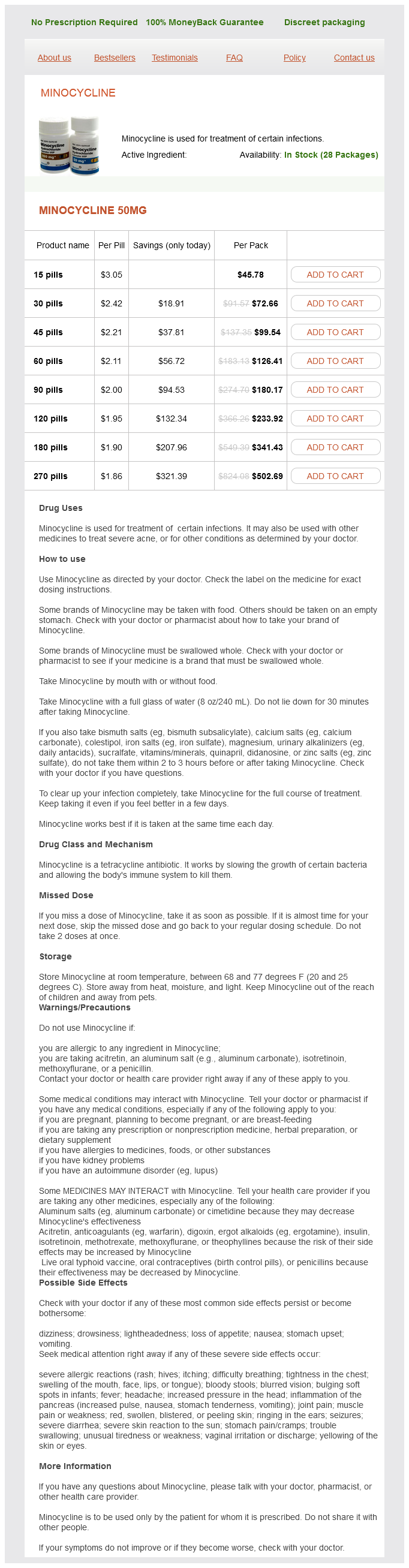
Minocycline 50mg
- 15 pills - $45.78
- 30 pills - $72.66
- 45 pills - $99.54
- 60 pills - $126.41
- 90 pills - $180.17
- 120 pills - $233.92
- 180 pills - $341.43
- 270 pills - $502.69
Enzyme histochemical demonstration of lactase and sucrase activity in resin sections: the influence of fixation and processing infection rate of ebola buy minocycline 50 mg with mastercard. Immunocytochemical demonstration of hormones in pancreatic and pituitary tissue embedded in methyl methacrylate. Superheating using pressure cooking: its use and application in unmasking antigens embedded in methyl methacrylate. Immunohistochemistry on resin embedded tissue for light microscopy: a novel postembedding procedure. A new acrylic resin formulation: a useful tool for histological, ultrastructural, and immunocytochemical investigations. The use of inert dehydration and glycol methacrylate embedding for immunogold localization of glomerular basement membrane components. An approach to post-embedding staining of protein (immunoglobulin) antigen embedded in plastic. Histochemistry and immunohistochemistry of fixation labile moieties in resin embedded tissue. Use of methacrylate de-embedding protocols for in-situ hybridization on semi thin plastic sections with multiple detection strategies. Horobin Introduction the physicochemical mechanisms of most histological stains are now understood. Detailed accounts and general overviews are to be found in the references and further reading at the end of this chapter. Histological staining methods from acid dyes to silver impregnation, involve broadly similar physicochemical principles. The present chapter aims to outline the major theories on common staining procedures and facilitate rational trouble-shooting if problems are encountered. Key questions to consider when seeking to understand histological stains are: · Why do any tissue components stain These questions can be answered for most stains, although some answers are complex. However, these methodologies are all influenced by selective uptake of stains and staining reagents into cells and tissues, and selective losses of stains from tissues. Which uptakes and losses occur depends both on binding equilibria and on rate factors. Nomenclature note: staining always involves the visual labeling of some biological entity by attaching, or depositing in its vicinity, a marker of characteristic color or form. In the physicochemical literature, to say a tissue component has a high affinity for a dye merely means there is a tendency for a stain to transfer from solution onto a section and this concept is used here. The magnitude of the affinity depends on every factor favoring or hindering this movement. The familiar stain-tissue attractions, including stainsolvent and stain-stain interactions, can be influential, as can solvent-solvent interactions. This account initially assumes staining reaches equilibrium, and the consequences of this not being reached are discussed later. The intensity of staining may also be influenced by stain solubility in solvent and tissue environments. However, as histologists and histochemists often emphasize reagent-tissue attractions as affinity sources, these interactions are discussed first. Reagent-tissue interactions Coulombic attractions have been termed salt links or electrostatic bonds, and have been the most widely 114 Why and how staining happens 115 Table 9. However, binding of dye ions to an ionic tissue substrate also depends on charge magnitude, the amount of nondye electrolyte in the dyebath, electrical repulsions between ions of similar charge, and swelling or shrinking of tissue substrates (Scott, 1973; Bennion & Horobin, 1974; Goldstein, & Horobin, 1974b; Horobin & Goldstein, 1974). Moreover, even uncharged tissue substrates acquire an ionic character after binding ionic reagents. This is also true for the large aromatic systems of bisazo dyes and bistetrazolium salts, halogenated dyes such as rose Bengal and phloxine, and indoxyl and naphthyl enzyme substrates (Horobin & Bennion, 1973). This attractive interaction arises when a hydrogen atom lies between two electronegative atoms. Water is hydrogen bonded extensively to itself, forming the clusters important for the hydrophobic effect discussed below. This effect also applies to molecules carrying hydrogen bonding groups present in many dyes and tissue components. As water molecules vastly outnumber dye molecules, hydrogen bonding is not usually important for stain-tissue affinity when aqueous solvents are used. Exceptions arise when hydrogen bonding is favored by the substrate, as with collagen tissue fibers (Prentø, 2007). This could explain strong staining seen with dyes such as eosin Y (4 arylbromo 116 9 Theory of histological staining Staining using Sudan dyes in non-aqueous solvents does not involve the hydrophobic effect. Adamson, 2012), the tendency of a system to change spontaneously to maximize its disorder, and for entropy to increase, provides an explanation. Presence of dye in solvent and lipid constitutes a more disordered system than dye restricted to the solution. Such increases in entropy involving substrate and stain occur in all types of histological staining. Covalent bonding between tissue and stain is yet another source of stain-tissue affinity.

ADDII BIOTECH is proud to announce that the company's manufacturing facility located at Baddii, Himachal Pradesh has recently been approved by Food & Drug Authority of Ghana.
Our Manufacturing capability includes a wide range of therapeutic products covering almost every segment.
ADDII BIOTECH given our strong emphasis on product quality and services.