
Mobic
General Information about Mobic
Mobic is prescribed for each short-term and long-term use, depending on the severity of the condition. It is often used to handle the signs of arthritis, including pain, swelling, and stiffness. However, it can also be prescribed for other circumstances such as menstrual cramps and acute ache.
Mobic just isn't appropriate for everybody, and it's essential to reveal any pre-existing health circumstances and drugs to a healthcare provider before starting treatment. Individuals with a historical past of heart, kidney, or liver disease, as properly as those taking other NSAIDs, blood thinners, or corticosteroids, is probably not prescribed Mobic or might require a special dosage.
In conclusion, Mobic is a extremely effective medication for managing the symptoms of arthritis and other conditions inflicting ache and irritation. Its longer period of action, security profile, and convenient once-daily dosage make it a most popular therapy possibility for many individuals. However, it's important to use Mobic as prescribed, disclose any pre-existing situations and drugs to a healthcare provider, and report any concerning unwanted facet effects. With the proper steering and monitoring, Mobic can provide relief and improve the quality of life for those affected by arthritis.
However, like some other medicine, Mobic additionally comes with potential unwanted side effects. The most common side effects include gastrointestinal signs similar to nausea, diarrhea, and abdomen pain. Rare however severe side effects embody liver and kidney issues, allergic reactions, and an increased danger of heart assault or stroke. Therefore, it is essential to seek the advice of a well being care provider earlier than beginning the treatment and to report any unusual unwanted effects whereas using Mobic.
Arthritis is a standard situation affecting hundreds of thousands of people worldwide. It is characterized by inflammation of the joints, which may result in pain, stiffness, and problem moving. There are multiple forms of arthritis, but the most typical forms are osteoarthritis and rheumatoid arthritis. Both of these circumstances can be debilitating and impact an individual's quality of life, making it essential to find effective treatments such as Mobic.
Additionally, Mobic has an extended period of action in comparability with other NSAIDs, making it a extra convenient possibility for those with continual circumstances. The commonplace dosage for Mobic is 7.5mg or 15mg once a day, which makes it a more manageable treatment possibility for patients, lowering the number of tablets they have to take on a daily basis.
One of the reasons Mobic is a preferred treatment for arthritis is its effectiveness in lowering pain. As an NSAID, it has a similar mechanism of action to aspirin, decreasing the production of prostaglandins, which are the chemicals liable for irritation and pain. By inhibiting their manufacturing, Mobic supplies relief from the symptoms of arthritis, allowing individuals to hold out their every day activities comfortably.
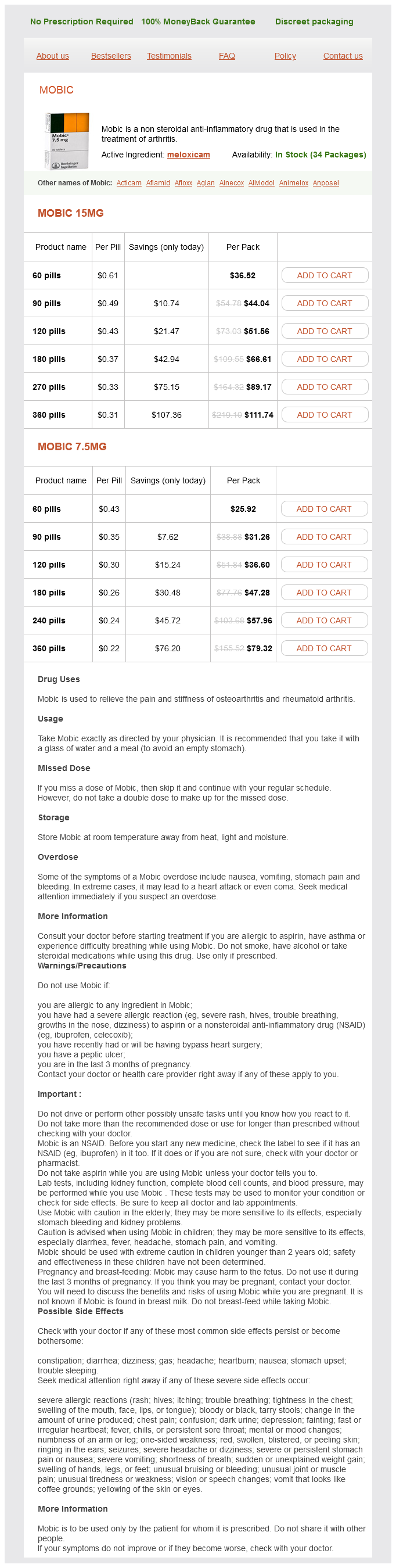
Mobic, also recognized as meloxicam, is a popular non-steroidal anti-inflammatory drug (NSAID) used to relieve pain and inflammation brought on by various situations, most commonly arthritis. It is a half of the family of drugs generally known as COX-2 inhibitors, which work by blocking the production of sure chemical substances in the physique that trigger ache and swelling.
Another benefit of Mobic is its security profile. It has been found to have lesser gastrointestinal unwanted effects in comparison with other NSAIDs, because it selectively targets COX-2 enzymes, that are responsible for irritation, while sparing COX-1 enzymes, which assist protect the abdomen lining. This makes it a more appropriate treatment for people who may be at risk for stomach issues.
The edema can have both a cardiogenic component end stage arthritis in dogs order mobic with american express, related to systemic hypertension and left ventricular dysfunction, and a neurogenic (pulmonary capillary leak) component. Life-threatening arrhythmias can occur in the setting of acute cerebrovascular disease. Ventricular tachycardia or fibrillation has been observed in patients with subarachnoid hemorrhage and head trauma. Stroke syndromes other than subarachnoid hemorrhage appear to be only rarely associated with serious ventricular tachycardias. Atrial arrhythmias including atrial fibrillation and regular supraventricular tachycardia have been observed. Atrial fibrillation is most common in patients presenting with acute thromboembolic stroke. Bradycardias including sinoatrial block, sinus arrest, and atrioventricular block occur in up to 10% of patients with subarachnoid hemorrhage. Treatment and Prognosis Beta blockers appear to be effective in decreasing myocardial damage and in controlling both supraventricular and ventricular arrhythmias associated with subarachnoid hemorrhage and head trauma. Beta blockers increase the likelihood of bradycardia and cannot be used in patients with hypotension requiring vasopressors. Life-threatening arrhythmias occur primarily in the first day after a neurologic event. Careful monitoring of potassium levels, especially in patients with subarachnoid hemorrhage, is warranted. Refractory ventricular arrhythmias have been controlled effectively with stellate ganglion blockade. Electrocardiographic abnormalities reflect unfavorable intracranial factors but do not appear to portend a poor cardiovascular outcome. The magnitude of peak troponin elevation is predictive for adverse patient outcomes including severe disability at hospital discharge and death. Head injury (blunt trauma or gunshot wound) and cerebrovascular accidents are the leading causes of brain death in patients being considered as heart donors. These donors can manifest electrocardiographic abnormalities, hemodynamic instability, and myocardial dysfunction related primarily to adrenergic storm and not to intrinsic cardiac disease. Experimental studies on whether contractile performance recovers with transplantation are still controversial. Optimization of volume status and inotropic support with careful echocardiographic evaluation and possibly left-heart catheterization can allow the use of some donor hearts that would have otherwise been rejected. The patient subsequently was treated with a beta-adrenergic blocker without further ventricular tachycardia. Adult cardiologists and electrophysiologists all participate in the management of patients. Controversies regarding appropriate use of pharmacotherapy and device therapy to manage cardiac manifestations in the neurologic diseases will be addressed further in clinical series and nonrandomized trials. The surety of therapeutic benefit afforded by assessment in a randomized controlled trial will not be available for a majority of these rare diseases. Gene-based or moleculartargeted therapy is under current evaluation in many of the neurologic diseases and holds future promise. Nigro V, Aurino S, Piluso G: Limb girdle muscular dystrophies: Update on genetic diagnosis and therapeutic approaches. Weidemann F, Rummey C, Bijnens B, et al: the heart in Friedreich ataxia: Definition of cardiomyopathy, disease severity, and correlation with neurological symptoms. Less Common Neuromuscular Diseases Associated with Cardiac Manifestations References the Muscular Dystrophies and Friedreich Ataxia 1. Rau F, Freyermuth F, Fugier C, et al: Misregulation of miR-1 processing is associated with heart defects in myotonic dystrophy. Wahbi K, Meune C, Porcher R, et al: Electrophysiological study with prophylactic pacing and survival in adults with myotonic dystrophy and conduction system disease. Berardo A, Musumeci O, Toscano A: Cardiological manifestations of mitochondrial respiratory chain disorders. Wahbi K, Larue S, Jardel C, et al: Cardiac involvement is frequent in patients with the m. Rudnik-Schoneborn S, Heller R, Berg C, et al: Congenital heart disease is a feature of severe infantile spinal muscular atrophy. Otten E, Asimaki A, Maass A, et al: Desmin mutations as a cause of right ventricular heart failure affect the intercalated disks. Suzuki S, Utsugisawa K, Yoshikawa H, et al: Autoimmune targets of heart and skeletal muscles in myasthenia gravis. Tung P, Kopelnik A, Banki N, et al: Predictors of neurocardiogenic injury after subarachnoid hemorrhage. The kidney has a central role in electrolyte balance, protein production and catabolism, and blood pressure regulation. Elucidation of these pathways has led to development of some of the key diagnostic and therapeutic targets in contemporary cardiovascular medicine. The CrCl is still used and preferred for renal drug dosing because it relies on actual body weight. Its low molecular mass and its high isoelectric point allow free filtration by the glomerulus and 100% reabsorption by the proximal tubule.
This article presents the differential diagnosis for optic neuropathies based on the optic disc appearance and discusses retinal disorders of particular interest in neurology arthritis glucosamine generic mobic 15 mg line. Chapter 45 discusses in more detail many of the entities described in this chapter. Once nerve fibers pass the lamina cribrosa, they are supported by oligodendrocytes and become myelinated. After exiting the orbit, the nerve enters the optic canal within the lesser sphenoid wing. In this space, the nerve is particularly vulnerable to trauma or compressive lesions (Prasad and Galetta, 2011). Orthograde transport (away from the ganglion cell body) occurs at two speeds: 400 mm per day for proteins and neurotransmitters packaged in vesicles, and 1 to 4 mm per day for structural elements of the cytoskeleton. The short posterior ciliary arteries provide blood supply to the optic nerve head and the subretinal choroid. Each posterior ciliary artery supplies a variable segmental territory of the optic nerve head, and because anastomoses in this blood supply are scant, it can suffer watershed ischemia during hypoperfusion (Hayreh and Zimmerman, 2007). Furthermore, the segmental blood supply underlies the sectoral disc swelling or atrophy that results from interrupted flow of a posterior ciliary artery and subsequent optic nerve infarction. In addition, papilledema commonly is bilateral, in contrast with other optic neuropathies including optic neuritis or nonarteritic ischemic optic neuropathy. Causes of pseudopapilledema include congenital anomalies, myelinated nerve fibers, and optic nerve head drusen (discussed later). On the other hand, only one-third of patients with optic neuritis will have optic disc swelling, and, when present, it is typically mild. Two main types of retinal ganglion cells exist: parasol cells (which project to the magnocellular layer and are specialized for motion perception and coarse stereopsis) and midget cells (which project to the parvocellular layer and are specialized for high spatial resolution, color vision, and fine stereopsis). The papillomacular bundle conveys axons from the fovea directly to the temporal margin (T) of the optic disc. The remainder of temporal ganglion cell axons is arranged in arcuate bundles above and below the fovea, arriving at the superior and inferior disc margins. B, Centrocecal scotoma (connecting to physiological blind spot) in a patient with optic neuropathy due to B12 deficiency. C, Superior altitudinal central scotoma in a patient with inferior optic nerve compression due to a meningioma. D, Inferior arcuate altitudinal scotoma in a patient with nonarteritic ischemic optic neuropathy. E, Superior altitudinal scotoma in a patient with arteritic ischemic optic neuropathy. Finally, compressive lesions may lead to chronic disc edema, optociliary shunt vessels, and glistening white bodies on the disc surface (pseudodrusen from extruded axoplasm). However, considerable overlap exists in the patterns of visual field loss caused by the different forms of optic neuropathy. Optic Neuritis Typical optic neuritis is an inflammatory optic neuropathy caused by demyelinating disease (Toosy et al. Visual loss in the affected eye typically occurs rapidly over several hours to a few days. Decreased color vision and contrast sensitivity are highly characteristic (Baier et al. In addition, pain with eye movements precedes the vision loss in approximately 90% of cases (Optic Neuritis Study Group, 1991). Note swelling and enhancement of the right optic nerve (black arrow) consistent with inflammation. Visual field defects commonly are present; they can be either diffuse or discrete scotomas and are nonspecific. Note mild nerve fiber layer swelling, greatest at the temporal portion of the disc (black arrow), without hemorrhages or cotton-wool spots. The prognosis for recovery of vision generally is good but is in relation to the severity of the initial deficit. It provides a reliable structural marker that complements clinical assessments of visual function. Because low-dose oral corticosteroids may be associated with an increased risk of recurrence of optic neuritis, this therapy should be avoided (Beck et al. Trials are ongoing to assess the efficacy of newer oral immunomodulatory agents to reduce the risk of relapse after an initial clinical presentation with optic neuritis. Thickness measurement is shown in black line, with normal range depicted in green zone; measurements within the yellow zone are borderline, and within the red zone are abnormally low. Plasmapheresis may be beneficial in the acute stage, and treatment with rituximab (a chemotherapeutic monoclonal antibody that depletes B cells) may be an effective disease-modifying agent (Wingerchuk et al. Note blurring of the nasal disc margin, with pallor and swelling of the nerve head (black arrow). The patient also has inner retinal ischemia evidenced by large cotton-wool spots (black asterisks). The condition is associated with polymyalgia rheumatica, consisting of proximal muscle ache, arthralgia, and stiffness, as well as with jaw claudication, fever, malaise, and scalp tenderness. The diagnosis is suggested by an elevated erythrocyte sedimentation rate and C-reactive protein and is confirmed by evidence of giant cells and endovascular inflammation on temporal artery biopsy. In approximately 25% of cases, vision is limited to hand motion perception or worse. Intravenous corticosteroids may help delay the progression of visual loss and decrease the likelihood of fellow eye involvement. The prognosis for recovery in the affected eye, however, is poor despite treatment. Other risk factors include a crowded optic nerve head and nocturnal hypotension, possibly precipitated by antihypertensive therapy (Arnold, 2003).
Mobic Dosage and Price
Mobic 15mg
- 60 pills - $36.52
- 90 pills - $44.04
- 120 pills - $51.56
- 180 pills - $66.61
- 270 pills - $89.17
- 360 pills - $111.74
Mobic 7.5mg
- 60 pills - $25.92
- 90 pills - $31.26
- 120 pills - $36.60
- 180 pills - $47.28
- 240 pills - $57.96
- 360 pills - $79.32
This effort has substantially advanced our understanding of both the underlying pathogenic mechanisms and epidemiology swollen joints in dogs front legs buy mobic 7.5 mg without a prescription. Current priorities include identification of patients most at risk and the development of preventive therapeutic strategies. Cardiovascular specialists should consider an underlying inflammatory disease in young patients with otherwise unexplained angina, myocardial infarction, or stroke. Patients with a rheumatic disease who suffer a myocardial infarction have worse outcomes in terms of both heart failure and mortality than does the age-matched general population. Traditional risk factors alone do not explain the increased burden of atherosclerosis, but inflammation may exacerbate the effects of classic risk factors. Various molecular mechanisms mediate the increased risk for atherosclerotic disease and cardiovascular events. Increased endothelial cell apoptosis and diminished capacity for repair may contribute. Clinical trials have demonstrated that this approach reduces symptoms and structural damage to joints. It remains uncertain, however, whether drugs that control synovitis also confer vascular protection. Infliximab therapy may improve endothelial function as measured by flowmediated dilation 4 to 12 weeks after infusion, whereas etanercept has been reported to reduce aortic stiffness. Data from the British Society of Rheumatology Biologics Register are more encouraging. Treatment of the arthritis must be combined with a careful review of classic risk factors and appropriate steps taken to modify these factors. The direct effect of chronic inflammation on vascular endothelium may itself promote atherogenesis, in addition to exacerbating the actions of traditional cardiovascular risk factors. Constitutional symptoms at initial evaluation include night sweats, lethargy, malaise, and weight loss. Frequent mucocutaneous features include the classic butterfly facial rash, oral ulcers, and alopecia. Hematologic involvement includes lymphopenia in most and frequently hemolytic anemia, neutropenia, and thrombocytopenia. A defect in the clearance of apoptotic cells results in the exposure of nuclear antigens to an immune system with hyperreactive B cells. Loss of immune tolerance results in the generation of autoantibodies and immune complexes. Deposition of immune complexes in target organs leads to activation of complement and tissue injury. Complement activation and consumption of C3 and C4 leading to reduced plasma levels characterize active disease. Cyclophosphamide and high-dose corticosteroids remain the first-line treatment of life-threatening complications, including myocarditis, cerebritis, severe hematologic involvement, and glomerulonephritis. A variety of regimens have been used, including combinations of rituximab, prednisone, and cyclophosphamide. Undertreated and/or persistently active disease has been associated with accelerated atherogenesis. Therefore adequate individualized immunosuppressive therapy should minimize cardiovascular complications. Aggressive management of traditional risk factors is also advocated, including diligent monitoring and tight blood pressure control. Caution should be exercised in patients with active myositis, however, because statin therapy can exacerbate this complication. The clinical data available do not support significant protection against atherosclerosis by statins 2 to 3 years after initiation, although longer-term analysis is awaited. In an inception cohort of 1249 patients monitored for 8 years, 97 vascular events were recorded, 31 of which resulted from atherosclerotic disease. The lipid-filled core and fibrous cap can be seen along with evidence of calcification. Subendocardial late gadolinium enhancement is present in the anteroseptal left ventricle (arrows) and extends from the base of the heart to the midventricular region, consistent with a previous subendocardial myocardial infarction. Fibrinoid necrosis, a specific feature of the medium- and small-vessel vasculitides, typically affects the inner layer of the tunica media. Complications include stenosis and occlusions resulting in organ ischemia, thrombosis, aneurysm formation, and hemorrhage. Thus diagnosis often depends on clinical findings, laboratory indices, and imaging. The immunopathogenesis of the vasculitides is complex, multifactorial, and poorly understood. Cardiovascular disease in patients with vasculitis, although relatively rare, can be life-threatening. Aortitis, hypertension, coronary arteritis, valvular heart disease, pericarditis, myocarditis, conduction abnormalities, accelerated atherosclerosis, and cardiac failure can all occur. This section focuses on the vasculitides most likely to be encountered by cardiovascular disease specialists. CardiovasCular disease and disorders of other organs Premature Atherosclerosis Systemic lupus erythematosus Rheumatoid arthritis Ankylosing spondylitis Psoriatic arthritis Gout Takayasu arteritis Giant cell arteritis Coronary Arteritis Systemic lupus erythematosus Takayasu arteritis Kawasaki disease Churg-Strauss syndrome Polyarteritis nodosa Granulomatous polyangiitis Rheumatoid arthritis Atherosclerosis in Association with Other Rheumatic Diseases the relationship between chronic inflammation and atherogenesis implies that many rheumatic diseases may be associated with premature and increased cardiovascular risk (Table 84-1). Because data in support of this hypothesis are derived from relatively small studies, important current clinical challenges include the need to determine (1) which rheumatic diseases pose the greatest cardiovascular threat, (2) a means of identifying subsets of patients most at risk, and (3) preventative strategies to minimize cardiovascular events.

ADDII BIOTECH is proud to announce that the company's manufacturing facility located at Baddii, Himachal Pradesh has recently been approved by Food & Drug Authority of Ghana.
Our Manufacturing capability includes a wide range of therapeutic products covering almost every segment.
ADDII BIOTECH given our strong emphasis on product quality and services.