
Monuvir
General Information about Monuvir
One of the key advantages of Monuvir is that it may be taken orally, making it easily accessible to a broader range of sufferers. This is in distinction to different COVID-19 treatments similar to remdesivir, which is run intravenously and requires hospitalization. Monuvir can additionally be being studied as a possible at-home therapy for people who have been exposed to the virus but have not yet developed symptoms. This could assist stop additional spread of the virus within households and communities.
Another concern is the potential development of drug resistance. As with any antiviral remedy, there is a possibility for the virus to mutate and turn out to be proof against Monuvir. However, the drug's builders are actively monitoring for any indicators of resistance and are additionally exploring combination therapies to counter this problem.
Monuvir is an oral antiviral drug developed by Merck and Ridgeback Biotherapeutics. It is a nucleoside analog and works by blocking the replication of the virus, thus stopping it from spreading throughout the body. This drug has been in improvement for years and was initially supposed for the remedy of influenza. However, because of its broad-spectrum antiviral activity, it was additionally tested to combat different viral infections corresponding to Ebola and now, COVID-19.
In December 2020, the FDA licensed and accredited the emergency use of Monuvir, also identified as Molnupiravir, as an oral antiviral therapy for COVID-19. This new treatment has generated lots of excitement and hope because it might potentially help fight towards the ongoing global pandemic. Let's take a better look at what Monuvir is and the means it could be used to fight COVID-19.
Another essential aspect of Monuvir is its potential to combat rising strains of the virus. Recent research have proven that it's efficient in opposition to the Delta variant, which is presently the dominant strain in plenty of countries. This is an encouraging signal, because the virus continues to mutate, and coverings want to find a way to keep up with these adjustments.
In conclusion, Monuvir exhibits promising leads to the battle against COVID-19. It has the potential to scale back the severity of signs and stop hospitalizations, making it a crucial software in managing the pandemic. However, it is important to continue following different preventive measures similar to carrying masks, social distancing, and getting vaccinated. As Monuvir becomes more broadly obtainable, it could probably be a useful addition to our arsenal within the battle in opposition to COVID-19.
In the clinical trial conducted by Merck, Monuvir showed promising ends in decreasing the viral load in sufferers inside five days of taking the drug. The trial additionally showed that patients who received the drug had a reduced probability of needing hospitalization or suffering from extreme signs. This was a significant breakthrough as there are at present no FDA-approved antiviral remedies for COVID-19.
Monuvir just isn't with out its limitations and challenges. It is only efficient within the early stages of COVID-19, and as the virus progresses, the drug's efficacy decreases. Therefore, it is crucial to start treatment as soon as possible after the onset of signs. Additionally, the drug has not been examined in pregnant ladies, and its safety and efficacy in this population are still unknown.
Its exact function is not known hiv infection malaysia order monuvir mastercard, but it is thought to provide a special chemo-sensory pathway of olfaction and affects the secretion of luteinizing hormone-releasing factor from the hypothalamus. They carry general sensations of pain, touch, and temperature from skin and proprioceptive sensations of vibration and muscle and joint sense. Functional Components A cranial nerve consists of motor fibres (motor nerve) or sensory fibres (sensory nerve) or both the motor and sensory fibres (mixed nerve). The primary sensory neurons of olfactory nerve lie on the body surface in the epithelial lining of the nasal cavity and their dendrites lie free in the mucous film. It is purely sensory and responsible for vision; hence, it is also called the nerve of sight. It is actually a tract of brain for it develops as an outgrowth of diencephalon during embryonic life. The primary sensory neurons of olfactory nerve (olfactory neurons) undergo continuous turnover, i. Functional Component Special somatic afferent fibres: They carry sense of sight from the visual field of the corresponding eye. Functional Components Special somatic afferent fibres: They carry special sensations of smell from the olfactory region of the nasal cavity and terminate in the olfactory bulb. Course, Relations, and Distribution Each olfactory nerve consists of about 20 minute bundles of non-myelinated nerve fibres. They arise from primary receptor neurons (modified bipolar neurons) of the olfactory epithelium of nasal cavity, pass through foramina in the cribriform plate of the ethmoid to enter the anterior cranial fossa, where they terminate in the olfactory bulb. The bundles of olfactory nerves are surrounded by three meninges (namely pia mater, arachnoid mater, and dura mater) near the cribriform plate. This provides a potential communication between the subarachnoid space and lymphatics of the nasal mucosa. Anosmia can occur for a number of reasons such as atrophic rhinitis (degenerative disorder of nasal mucosa), fracture of the anterior cranial fossa (ethmoidal fracture), etc. Course and Relations the fibres of optic nerve arise from ganglion cells (secondorder neurons) in the neural layer of the retina of the eyeball, converge toward the optic disc at the posterior pole of the eyeball, pierce the outer layer of retina, choroid, and sclera to leave the eyeball. Immediately after emerging from the eyeball, the fibres unite to form the optic nerve, which passes posteromedially through the posterior half of the orbit and enters the middle cranial fossa through the optic canal. In the middle cranial fossa, optic nerves of two sides unite to form the optic chiasma. The midregion of optic chiasma is composed of crossed fibres from the medial/nasal halves of the retina of both eyes, while the lateral region is made up of fibres from the lateral/temporal half of the retina of the ipsilateral eye. The third-order neurons arise in the lateral geniculate body, run in the retrolenticular part of the internal capsule, and form optic radiations. The fibres of optic radiation terminate in and around the calcarine sulcus of the occipital lobe (visual cortex). Some of the fibres from the lateral geniculate body reach the pretectal area of the midbrain and form a part of the pathway for light reflex. It is divided into three parts: (a) intraorbital part, (b) canalicular part, and (c) intracranial part. General visceral efferent fibres: They supply the sphincter pupillae and ciliaris muscles. They arise from the parasympathetic component of oculomotor nucleus (also called the EdingerÂWestphal nucleus). The postganglionic parasympathetic fibres arise from the ganglion and supply the sphincter pupillae and ciliaris muscles. It runs forward and laterally between the posterior cerebral and superior cerebellar arteries and lateral to the posterior communicating artery, passes through the tentorial notch of tentorium cerebelli to reach the middle cranial fossa. Here it pierces the dura mater in the oculomotor triangle lying in between the free and attached margins of tentorium cerebelli in the roof of the cavernous sinus and enters the lateral wall of the cavernous sinus where it lies superior to the trochlear, ophthalmic, and maxillary nerves, and lateral to the internal carotid artery. In the anterior part of the cavernous sinus, the nerve divides into upper and lower divisions. The two divisions enter the orbit through the superior orbital fissure within the common tendinous ring. The smaller upper division passes above the optic nerve on the inferior surface of superior rectus (which it supplies), and then passes through the superior rectus to supply the levator palpebrae superioris. The larger interior division of the oculomotor nerve passes below the optic nerve and immediately gives three branches which supply the medial rectus, inferior rectus, and inferior oblique muscles. The nerve to inferior oblique gives motor root (parasympathetic root) to the ciliary ganglion located in the posterior part of the orbit. The postganglionic fibres from this ganglion run through short ciliary nerves and supply the sphincter pupillae and ciliaris muscles. Clinical correlation Lesions of oculomotor nerve: the damage of oculomotor nerve may occur due to the following factors: (a) Compression by aneurysm of the posterior communicating artery as it passes between posterior cerebral and superior cerebellar arteries. The damage of oculomotor nerve clinically presents as: · Ptosis (drooping of the upper eyelid), due to paralysis of the levator palpebrae superioris. It is purely motor and supplies only one muscle - the superior oblique muscle of the eyeball. It is the only cranial nerve whose nuclear fibres decussate before emerging on the surface of the brain. Functional Components and Nuclei General somatic efferent fibres: They arise from the trochlear nucleus in the midbrain and supply the superior oblique muscle of the eyeball.
Sphenomandibular ligament: It is attached above to the spine of the sphenoid and below to the lingula and lower margin of the mandibular foramen of the mandible antiviral rx order monuvir with amex. Medially, it is related to: (a) medial pterygoid, (b) chorda tympani nerve and (c) wall of the pharynx. Near its lower end the sphenomandibular ligament is pierced by mylohyoid nerve and vessels. Clinical correlation the sphenomandibular ligament is an important landmark for administration of local anesthetic during inferior alveolar nerve block. Auriculotemporal nerve: Its articular twigs enter the joint from its posterior aspect. The stylomandibular ligament is formed due to thickening of the investing layer of deep cervical fascia, which separates the parotid and submandibular glands. In occlusion, the teeth themselves stabilize the mandible on maxilla and no strain is thrown on the joints when an upward blow is received on the mandible. Further in the occluded position, the forward movement of condyle is discouraged by the articular eminence and by the contraction of the posterior fibres of the temporalis muscle, while the backward movement of the condyle is prevented by the lateral ligament and the contraction of the lateral pterygoid muscle. Posterior: (a) Postglenoid part of parotid gland separating it from external auditory meatus. The lower jaw can be depressed, elevated, protruded retracted and moved from side-to-side, by movements at temporomandibular joints. The lower menisco-temporal compartment permits rotation around two axes (a) a transverse axis, during depression and elevation and (b) a vertical axis during side-to-side/chewing movements. Depression Elevation Protraction Retraction Side to side (Chewing) movements of suprahyoid muscles, viz. Elevation (elevating of jaw to close the mouth): During elevation the movements take place in a reverse order to that of depression, i. Protrusion/Protraction: During this movement, mandibular teeth move forward in front of maxillary teeth. In this act, head of mandible along with articular disc glide forwards in the upper meniscotemporal compartment on both sides by simultaneous action of medial and lateral pterygoids of both sides. Retraction: During this movement, the head of mandible along with articular disc glide backwards in the upper meniscotemporal compartment by the contraction of the posterior fibres of temporalis muscle and bring the joint in the resting position. The forceful retraction is assisted by deep fibres of masseter, digastric, and geniohyoid muscles. At the end of this movement the head of the mandible comes to lie underneath the articular tubercle. Side-to-side (Chewing) movements: these movements occur alternately in the right and left temporomandibular joints. In chewing movements, the head of the mandible on one side glides forwards along with the disc (as in protraction), but the head of the mandible on the opposite side merely rotates on the vertical axis. During this movement, the medial and lateral pterygoids of one side contract alternatively with those of opposite sides. The alternate movements of this kind on the two sides result in side-to-side movements of the lower jaw. Muscles Producing Movements Depression (Opening of Mouth) It is produced mainly by lateral pterygoid helped by gravity. The digastric, geniohyoid, and mylohyoid muscles help when the mouth is opened widely or against resistance. Elevation (Closing the Mouth) It is caused by medial pterygoid, masseter, and temporalis (vertical fibres). Therefore, when attacked by a street dog, it is advisable to keep the mouth of dog closed, if possible. Depression (lowering of jaw to open mouth): During depression, the head of mandible along with an articular disc glide forward in the upper meniscotemporal compartment on both the sides by the contraction of lateral pterygoid muscle. At the same time head rotates forward underneath the articular disc by the contraction 148 Textbook of Anatomy: Head, Neck, and Brain Protraction It is done by lateral and medial pterygoids and masseter. It may be assisted by middle and deep fibres of the masseter, the digastric and geniohyoid muscles. Side-to-side (Chewing) Movements these movements are performed by alternate contraction of medial and lateral pterygoids on each side. Movements of the temporomandibular joint and muscles producing them are summarized in Table 10. Elevation Protrusion (Protraction) Retraction Side-to-side (chewing) movement Clinical correlation · Palpation of the temporomandibular joint and associated muscles: the bilateral palpation is must to assess the entire joint and its associated muscles. Then he is asked to move the opened jaw to left, and to right, and finally he is asked to move the jaw forward. For digital palpation of condyle of moving mandible place a finger into the outer portion of the external auditory meatus. When the mouth is open, the mandibular condyles move forward and lie underneath the articular eminences. In this position, if there is excessive opening of mouth as during yawning, sudden violence or convulsive spasm of lateral pterygoid muscles, the head of mandible of one or both sides may slip anteriorly and get locked into the infratemporal fossa; as a result the mouth cannot be closed anymore and any passive effort to do that will invariably fracture the neck of the mandible on one or both sides. To reduce dislocation, the condyle must be lowered and pushed back behind the summit of articular eminence into the articular fossa. Thus the reduction is done by depressing the jaw with thumb placed on the last molar teeth, and simultaneously elevating the chin. The typical presenting symptoms are:  Diffuse facial pain, due to spasm of masseter muscle. It occurs when the posterior attachment of the disc becomes stretched or detached, allowing the disc to become temporarily or permanently trapped anteriorly. Principal muscles (a) Temporalis (b) Masseter (c) Lateral pterygoid (d) Medial pterygoid. Principal Muscles of Mastication the characteristic features of the principal muscles of mastication are as follows: 1.
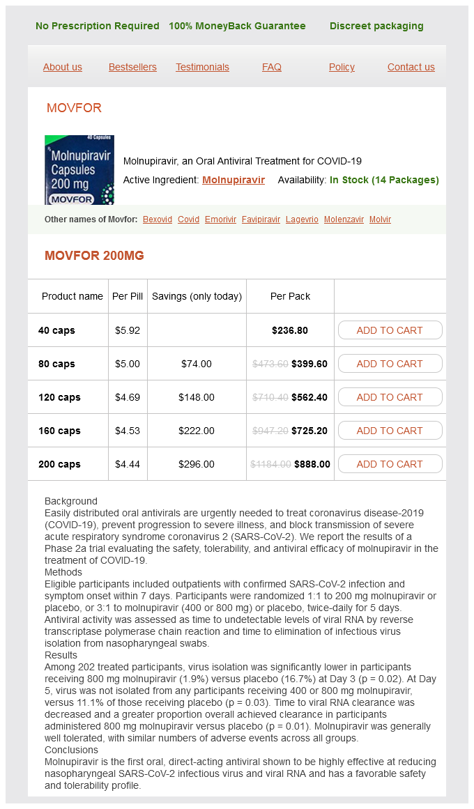
Monuvir Dosage and Price
Movfor 200mg
- 40 caps - $236.80
- 80 caps - $399.60
- 120 caps - $562.40
- 160 caps - $725.20
- 200 caps - $888.00
The decussation of the superior cerebellar peduncles occupies the central part of the tegmentum and forms the most important feature in the lower part of the midbrain antiviral for chickenpox discount monuvir online american express. The lemnisci are arranged in the form of a curved compact band of white fibres in the ventrolateral part of the tegmentum, lateral to cerebellar decussation and dorsal to the substantia nigra. From medial to lateral side, these are: medial lemniscus, trigeminal lemniscus, spinal lemniscus, and lateral lemniscus. The central grey matter in each half contains two nuclei: the oculomotor nerve nucleus and the mesencephalic nucleus. The nuclei of two sides fuse together forming a single complex having a triangular outline. The emerging fibres of oculomotor nerve pass ventrally through the tegmentum, intersecting red nucleus and medial part of the substantia nigra and emerge in the posterior part of the interpeduncular fossa through the sulcus on the medial aspect of crus cerebri. The nucleus of superior colliculus consists of cells which are involved in general light reflexes. Pretectal nucleus is a small group of nerve cells and lies deep to the superolateral part of the superior colliculus. Red nucleus is a cigar-shaped mass of grey matter which appears ovoid in cross section. It is situated in the tegmentum, ventral to the 3rd nerve nucleus and dorsomedial to the substantia nigra. Decussation of fibres (tectospinal and tectobulbar tracts) arising from superior colliculi forming dorsal tegmental decussation (of Meynert). Decussation of fibres (rubrospinal tracts) arising from red nuclei forming ventral tegmental decussation (of Forel). It is characterized by the loss of upward gaze without affecting the other eye movements. The cranial end of the neural tube forms the brain, while the caudal end forms the spinal cord. The nerve cells in the wall of the neural tube arrange themselves into functional groups. Medial Longitudinal Fasciculus Medial longitudinal fasciculus is a heavily myelinated composite tract found in the paramedian plane of the brainstem. The dorsal and ventral laminae are also frequently termed alar and basal laminae, respectively. The alar lamina consists of sensory cells and basal lamina consists of motor cells. The cells in each lamina are arranged into two columns: the one near the sulcus limitans, concerned with the innervation of viscera, is called the visceral column and the one away from sulcus limitans, concerned with innervation of somatic structures, is called the somatic column. The characteristic features of this syndrome are as follows:  Ipsilateral lateral squint, due to involvement of the 3rd cranial nerve. It is characterized by the following signs and symptoms:  Ipsilateral lateral squint and ptosis, due to involvement of oculomotor nerve fibres. Apart from this, an extra special somatic column appears in the most lateral part of the alar lamina to innervate the vestibular and cochlear apparatuses. As the development proceeds, each column differentiates into two or more discrete cranial nerve nuclei (Table 24. Reticular formation extends cranially to the diencephalon and caudally to the spinal cord. The reticular formation receives data from most of the sensory systems of the body and relay them to all the levels of the neuraxis. It is important for the maintenance of sleep wake cycle, level of consciousness, and alertness or mutism. Although reticular formation is described as a network of nerve fibres intermingled with nerve cells, on careful examination it reveals fairly localized cell groups called reticular nuclei in certain regions. The reticular pathways are polysynaptic, both ascending and descending, and crossed and uncrossed. Clinical correlation the visual and acoustic stimuli can stimulate the reticular activating system and thus maintain alertness and attention. For this reason, the stimuli such as sound of ringing alarm clock or sudden bright light can arouse consciousness. He also noted that when he walks, his left arm remains flexed and his left leg remains extended. Further, he has to swing his left lower limb outward to avoid dragging the foot on the ground. On examination, the neurologist found: (i) no signs of facial palsy, (ii) deviation of tongue to the right side on protrusion, (iii) atrophy of tongue on the right side, and (iv) loss of position and vibration sense on the left side. It is located in the posterior cranial fossa underneath the tentorium cerebelli and behind the pons and medulla oblongata. In general, the fissures and folia of the cerebellum lie transversely from side-to-side across the whole extent of the cerebellum. Each hemisphere of the cerebellum is connected to three parts of the brainstem by three pairs of the large fibre tracts called cerebellar peduncles. The cerebellar disease is manifested by motor disturbances, including inability to stand upright, staggering gait, hypotonia, and failure of coordination. The superior and inferior aspects of vermis are termed superior and inferior vermis, respectively. The ridge-like superior vermis is continuous on either side with the superior surface of cerebellar hemisphere imperceptively. The inferior vermis is more clearly demarcated from the hemispheres in the floor of vallecula cerebelli. The inferior surface presents a deep median notch called vallecula which separates the two cerebellar hemispheres.

ADDII BIOTECH is proud to announce that the company's manufacturing facility located at Baddii, Himachal Pradesh has recently been approved by Food & Drug Authority of Ghana.
Our Manufacturing capability includes a wide range of therapeutic products covering almost every segment.
ADDII BIOTECH given our strong emphasis on product quality and services.