
Rumalaya gel
General Information about Rumalaya gel
In addition to its pain-relieving properties, Rumalaya gel additionally has anti-inflammatory effects. This makes it not solely efficient for treating ache but also for reducing swelling and stiffness associated with joint and muscle problems. Regular use of the gel can also help improve mobility and range of movement in affected areas, making it an excellent option for those suffering from chronic situations like arthritis.
Moreover, Rumalaya gel is a non-greasy method, not like many other topical ache reduction products. Its non-greasy nature makes it straightforward to make use of on any a part of the physique without leaving any residue or staining on garments. This makes it an ideal alternative for many who need to use it during the day, at work, or while partaking in bodily activities.
The main active components in Rumalaya gel are Indian Winter Green (Gandhapura taila) and Boswellia (Shallaki). The Indian Winter Green is a natural ache reliever that works by blocking the production of inflammatory mediators and reducing ache and irritation. Boswellia, however, has anti-inflammatory and analgesic properties and helps in decreasing swelling and pain related to joint and muscle disorders.
One of the most important advantages of using Rumalaya gel is its fast motion. Many customers have reported feeling aid inside minutes of applying the gel to the affected space. This is as a outcome of the gel is designed to penetrate deep into the skin and attain the affected tissues and muscle tissue, offering fast and efficient ache relief. The gel also has a pleasing cooling impact, providing a soothing sensation to the affected space.
What units Rumalaya gel apart from different topical pain aid products is its natural formulation. The gel is created from all-natural ingredients, making it a secure and efficient possibility for these looking for a natural remedy for their ache. Unlike prescription medicines, Rumalaya gel does not have any antagonistic unwanted side effects and can be utilized for prolonged durations without any harm.
This gel is a herbal formulation that's made up of pure ingredients and is efficient in treating varied musculoskeletal circumstances like arthritis, osteoarthritis, rheumatoid arthritis, sprains, strains, and other joint and muscle pains.
In conclusion, Rumalaya gel is a potent topical utility that gives quick and efficient reduction from ache. Its herbal formulation, quick action, non-greasy formula, and anti-inflammatory effects make it a popular alternative among these seeking a natural and protected resolution for joint and muscle pain. Whether you would possibly be an athlete, an office worker, or somebody suffering from chronic pain, Rumalaya gel is a must have in your medication cabinet for quick and long-lasting ache relief.
Aside from its quick action, another advantage of Rumalaya gel is that it's easy to make use of and does not require any particular skills or equipment. The gel comes in a convenient tube that makes it straightforward to apply on to the affected area. Simply squeeze a small amount onto your fingertips and gently therapeutic massage it into the skin until totally absorbed. The gel can be used up to three to 4 occasions a day depending on the severity of the ache.
Difficulties in meeting primary efficacy endpoints in Pivotal clinical studies can often be attributed to missing one of these enabling features spasms synonyms rumalaya gel 30 gr order with amex. Anatomy enables simple, minimally invasive surgical procedure the vagus nerve has become a popular target for autonomic neuromodulation therapies. At the mid-cervical level the vagus nerve lies superficially within the carotid sheath. The vagus contains pathways to and from all the major organs in the viscera, and regulates critical systems implicated in numerous diseases and disorders such as the inflammatory reflex, digestion, and bronchoconstriction. A major advantage of targeting the vagus nerve at the mid-cervical level is that this location is typically directly visible via ultrasound, and its location is consistent relative to the carotid arteries, which are also visible. This surgical complexity may have led to an increased number of adverse events such as nerve damage-related paresthesias. One major advantage of the tibial nerve is that it is large enough to visualize via ultrasound, a critically enabling facet of a needle-stick procedure. It should be noted that a minimally invasive surgical procedure such as a percutaneous needle stick does not guarantee fewer adverse events. Although the electrode and lead designs for the StimWave and StimGuard system enable wireless power and telemetry, the size and configuration of the implanted electrode arrays are similar to existing systems powered through a wired connection to an implantable pulse generator. SetPoint Medical (inflammation) and Microtransponder (tinnitus, stroke rehabilitation), amongst others, are developing technology to reduce the size of the implantable electrode and transceiver to stimulate points along the vagus nerve. The end-goal is to reduce the size of the full implant- including electrodes and transceiver for power and telemetry - to fit within an injectable hermetic enclosure the size of a grain of rice. Although there has been considerable interest in developing vascularly implanted miniaturized wireless systems for indications other than cardiac pacing, developing a simple-to-implant device for other indications with the necessary focality of stimulation from within or across a vessel wall has remained elusive. Activation of target nerves is separable from off-target nerves There is a trade-off between a minimally invasive electrode design and an electrode design that can minimize the activation of off-target nerves and muscles that lead to side-effects that can limit the therapeutic efficacy. These designs allow the wire to be implanted percutaneously through a needle or introducer, and the barb provides anchoring to maintain the position of the electrode once the introducer is retracted. Unfortunately, this simple design is not viable for many nerve stimulation applications as the applied current can create unwanted activation of nearby untargeted nerves or muscles at subthreshold levels for intended therapeutic stimulation of target nerves. Programming procedures that are driven first and foremost by limiting side effects do not guarantee the device is engaging the intended neural target, especially when biomarkers of target engagement are often limited or unreliable. The standard vagal nerve stimulator, for example, utilizes an insulating cuff that is wrapped around the cervical vagus in conjunction with a tripolar electrode design to limit the spread of current outside of the insulating cuff. Uninsulated electrodes similar to the Permaloc design that can be implanted via a needle injection have been attempted for vagal nerve stimulation in animals, but have been critically limited by unintended activation of the neck muscles at levels still too low for therapeutic activation of the vagus. A number of commonly used techniques to maximize intended effect while minimizing unintended effects are critically dependent on the functional anatomy of the neural target and surrounding tissue. Consequently, targets with functional anatomy where the stimulating electrode can be consistently placed and maintained closer to target nerves are preferred. There is also considerable interest in utilizing multiple cathode and anode electrode configurations to steer current to preferentially activate tissue between electrode contacts instead of at the location of the electrode. Although many market approved devices are capable of current steering, the ability of these techniques to create virtual electrodes for consistent preferential activation between electrode locations in long-term implants remains unproven. Therefore, the ability to predict and maintain a virtual cathode or anode for preferential activation of tissue to enhance desired effects and prevent unwanted side effects may be a valuable but limited tool. A second common technique is manual calibration of the energy applied (pulse width and pulse amplitude) to find a setting that directly initiates action potentials in the fibers of interest while remaining sub-threshold for activating off-target fibers. Consequently, implant locations where there are clear morphological differences between target fibers and off-target fibers that can be differentiated through programming are preferred. Some systems have experimented with intermittent stimulation, such as stimulating one minute out of every five, but primarily to save battery life. Modern systems, however, are increasingly attempting to decipher and imitate the natural patterning of signals sent by nerve fibers to drive desired outcomes. For example, the BioControls system monitors the cardiac cycle to deliver stimulation in rhythm with the intrinsic beating of the heart, in order to mimic the rhythm of natural signals sent by sensory afferents from the heart during normal function. Whether more natural temporal patterning can enhance target effect while minimizing offtarget effects compared to stimulation at a single fixed frequency in human patients remains unclear. SetPoint Medical is currently exploring this idea using existing vagal nerve stimulators in exploratory clinical trials in Europe. Similarly closed-loop systems such as the NeuroPace Responsive Neural the Autonomic Nervous System 33 Stimulation system use sensors to stimulate only when an active seizure is about to occur to minimize side effects and maximize battery life. Therapies in which stimulation only needs to be administered for a few minutes a day could potentially enable a host of less specific non-invasive neuromodulation techniques, as offtarget effects that would be intolerable for a continuous therapy may be easily manageable for short periods. Low variability in functional anatomy from patient to patient Given a lack of high resolution tools for clinical research, variability in functional anatomy is difficult to assess from patient to patient, but has increasingly been implicated as a contributor to highly variable functional outcomes from patient to patient evident with many neuromodulation therapies. Although medical adherence and type of therapy administered as a function of race and cultural norms may have strongly contributed to this disparate result, genetic variability is also postulated as a root cause. Similarly, very little is known about the variability in the organization of fibers from patient to patient in popular implant locations like the cervical vagus. The cervical vagus consists of approximately 100,000 fibers, of which 65-80 percent are unmyelinated visceral afferent sensory fibers from a multitude of visceral organs. The distance between afferents and efferents to and from a specific organ and the epineural cuff electrode at the level of the cervical vagus is largely unknown, and may vary greatly across a population. Consequently, there is increasing interest in therapeutic targets with consistent functional organization of both intended and unintended neural targets from patient to patient, in order to develop a single therapy that maximizes the number of responsive patients.
Better compensation occurs when gain is increased muscle relaxant egypt rumalaya gel 30 gr, but that risks making the system unstable. Positive Feedback Negative feedback becomes positive when the feedback is added to , rather than subtracted from, the input, so that "error" growth accelerates. The resulting explosive growth of output can produce useful power surges and rapid state transitions, but, to be useful, these rapid changes must be controlled. Positive feedback is often controlled by nesting it within a larger negative feedback system that limits total change. For example, the rapid membrane depolarization of an action potential is produced by the positive feedback between depolarization and sodium channel opening. In locomotion, positive feedback assistance reflexes promote rapid transitions between stance and swing that are stabilized by negative feedback. At the end of stance, a leg perturbation or an attempt to lift it may trigger an assistance reflex that excites leg flexor motor neurons. The assistance reflex may increase as the leg rises and be extinguished as the leg reaches the limits of elevation (Chung et al. A cockroach walking at high speed over a rough terrain, crayfish wrestling for social dominance, and a praying mantis carefully stalking its prey before launching a directed attack, all require a high degree of coordination between brain centers, thoracic motor centers, and multiple streams of sensory feedback continuously arriving from the periphery. Posture and locomotion are controlled in arthropods using sensory feedback from a wide variety of sensors and musculoskeletal structures and the control principles and organizational patterns described above. We therefore here provide only a brief summary to acquaint readers unfamiliar with the general organization. Muscles originate from interior points of the exoskeleton and project across a joint to attach on the cuticle or an apodeme, an internal tendon-like structure. Insect and crustacean legs are segmented tubular structures that emerge from the thorax, itself a fused multi-segmental structure. The legs contain five (insects) or seven (decapod crustaceans) serially arranged segments, ending with single or paired (pincer) claws. The leg segments are linked together by bicondylar hinge joints that allow the claw to move through a wide range of the space around the body. Like spindles, they receive efferent innervation that enables them to play a similar role in feedback control (Head and Bush 1991). Tendon receptors, like Golgi tendon organs, respond to apodeme force (Hartman 1985). Hair-like receptors respond to contact with the ground, with another body segment at a joint, or with an external object or force on the leg. Each bilaterally symmetric thoracic ganglion controls a pair of legs and contains about 2,000 neurons, 1,900 of which are interneurons (Wiersma 1961). Sensory afferents project to their segmental hemi-ganglion and motorneurons project to their segmental muscles. While the distal portion of the leg is supplied by hundreds of sensory afferents from cuticular hairs (Marchand et al. Unlike vertebrates, arthropods possess inhibitory motorneurons that prevent excitation of a muscle or speed muscle relaxation (Govind and Atwood 1982). One inhibitory motorneuron, the common inhibitor, innervates all the muscles of a leg. Neurons in the central complex of the insect brain are important for decisions related to walking speed and movement around barriers (Bender et al. These networks presumably translate unpatterned descending "commands" into patterns of coordinated leg movement, although the details are unknown. Nonetheless, it is clear that static postures are maintained through negative feedback resistance reflexes in which nearly 276 Neurobiology of Motor Control: Fundamental Concepts and New Directions all the proprioceptors and exteroceptors of the leg participate. As in insects and vertebrates, the excitatory resistance reflexes are monosynaptic and the inhibitory reflexes are di- or polysynaptic. Many resistance reflexes span more than one joint, so that the leg movements are directed in more than a single plane. Polysynaptic reflex responses in locusts are produced by afferents that excite non-spiking interneurons with wide effects in the thoracic ganglion (Burrows et al. Reflexes evoked by single proprioceptors or exteroceptors are thus part of a complex, but poorly understood, postural control system that works to stabilize posture through many parallel negative feedback pathways. Nervous systems exist in "quiescent" states where rhythmic motor patterns are inactive and resistance reflex responses are the rule, and "active" states characterized by at least some rhythmic motor activity and active resistance and assistance reflex responses (Chung et al. Rhythmically activated resistance reflexes can produce rhythmic motor patterns in quiescent nervous systems, but these are readily seen to be artifactual. Thus, if these resistance reflexes occurred in a walking animal, they would oppose the movement. This does not happen in active preparations or during walking, indicating that such unwanted resistance reflexes are prevented from occurring when the nervous system is in the active state. Pre-synaptic inhibition targets the afferent-to-interneuron connection that mediates the reflex, and often spares the outputs of the same afferents to other targets and the responses of the motor neurons to other inputs. Strong pre-synaptic inputs can also sufficiently depolarize the pre-synaptic terminal to evoke spikes that travel antidromically to the distal spike generating mechanism of the afferent. Antidromic spikes can inhibit the spike generating mechanism for hundreds of milliseconds, thereby preventing afferent participation in all reflex responses (Cattaert et al. During the early part of stance, when the leg is firmly planted and bearing a load, a resistance reflex to upward perturbation of the leg would be helpful. Reflex reversal also occurs at other joints in the same leg upon transition from a quiescent to an active state.
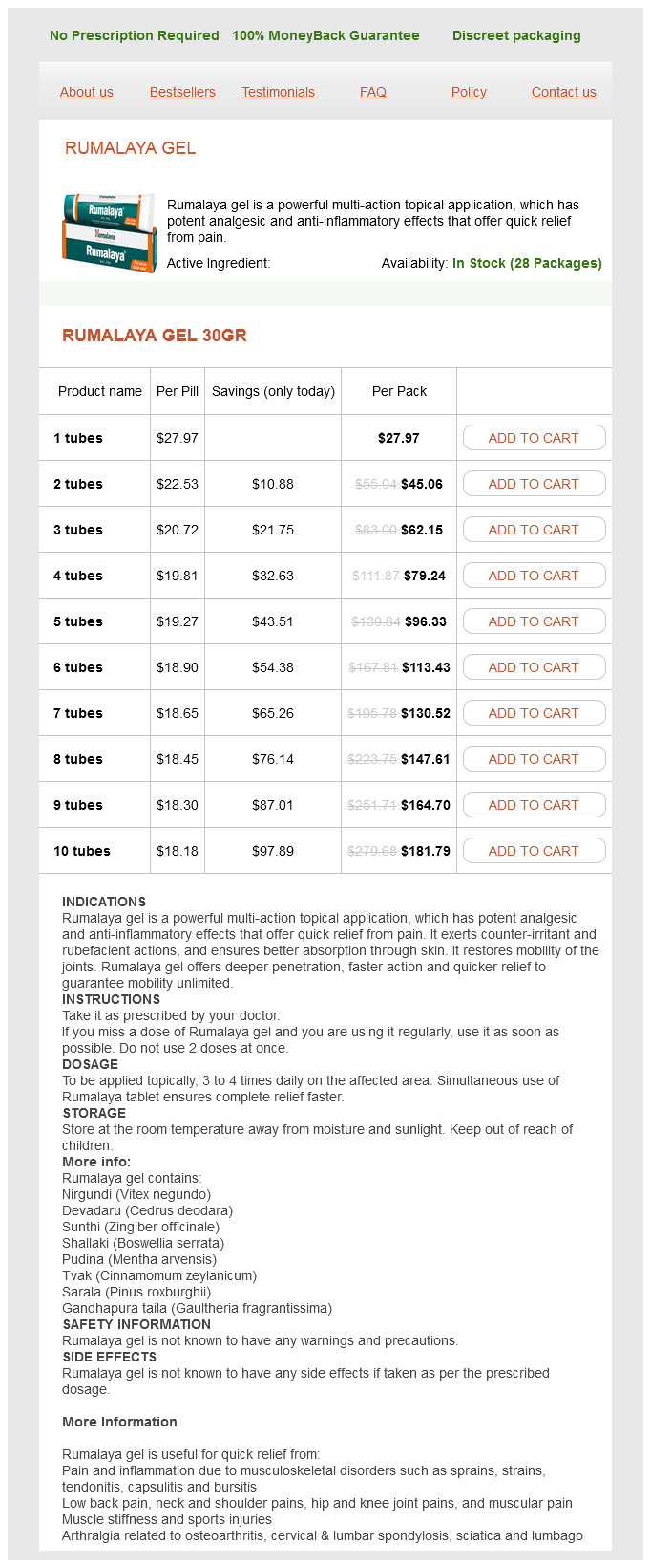
Rumalaya gel Dosage and Price
Rumalaya gel 30gr
- 1 tubes - $27.97
- 2 tubes - $45.06
- 3 tubes - $62.15
- 4 tubes - $79.24
- 5 tubes - $96.33
- 6 tubes - $113.43
- 7 tubes - $130.52
- 8 tubes - $147.61
- 9 tubes - $164.70
- 10 tubes - $181.79
It is important to note that the injection techniques included in all those nine studies were all "blind" injection with the exception of one study muscle relaxant 8667 rumalaya gel 30 gr order mastercard, in which x-ray confirmation was performed in a portion of the patients. A recent large pragmatic randomized controlled trial showed that the subacromial steroid injection improved pain and functional outcome at 1 and 6 weeks and there was no difference compared with exercise alone at 3 and 6 months. The anterior portion of the deltoid muscle was reflected to show the underlying rotator cuff muscle. Ultrasound-Guided Injection Technique the patient can be placed either in the supine or sitting position with the ipsilateral arm in the Middleton or modified Crass position (the palmar surface of the hand touching the buttock). The posterior portion of the deltoid muscle was partially removed to show the underlying muscle. Patients with biceps tendinitis present with anterior shoulder pain and tenderness over the bicipital grove. The insert on the left shows the position of the patient and the ultrasound probe, and the one on the right shows the probe and the structures underneath. Note that the portion of the supraspinatus tendon lateral to the acromion process was significantly increased by this maneuver. Note that the medial end of the ultrasound probe is place over the acromion (Acr). However, in a subject with a slim body build, the probe can be placed in another orientation, as shown in the right insert. The ultrasound image on the left shows the needle (arrow heads) inserted with in-plane technique to the subacromial subdeltoid bursa, as highlighted by the peribursal fat (line arrows). The image on the right shows the presence of local anesthetic following the injection, with the separation of the deltoid muscle and supraspinatus tendon. The insert shows the position of the probe and the corresponding anatomical structures underneath. The local anesthetic is seen surrounding the bicep tendons in the bicipital groove (line arrows). The complex movement, shear stress, and high compressive force from the surrounding muscles contribute to the degenerative process of the joint. The inferior surface of the joint is in direct contact with the subacromial bursa and supraspinatus muscle and may play a role in the development of the impingement syndrome. Ultrasound-Guided Injection Technique the patient position can be either in sitting (chair with back support) or supine position, with the arm in the neutral position, as the deep joint space is the widest at this position. In a young, healthy subject, a fibrocartilaginous disc is usually seen as a slightly hyperechoic wedge-shaped structure attached to the superior joint capsule and is mobile in its location depending on the arm position. The superior acromioclavicular ligament is a hyperechoic fibrillar structure abutting the bony surfaces of the joint. The upper insert shows the position of the probe and the patient, and the lower insert shows the position of the probe and the structures underneath. The corresponding ultrasound image shows the acromioclavicular joint with the image of the needle (solid arrow). The volume of injectate is usually 2 mL, and a successful injection is indicated by the elevation of the capsule and widening of the joint space under real-time scanning. Conclusion Ultrasonography is a very useful tool allowing accurate localization of the various target structures for shoulder injections and real-time guidance of the needle insertion. A good understanding of the anatomy and sonoanatomy of the shoulder is of paramount importance in performing the ultrasound-guided injections. Estimating the burden of musculoskeletal disorders in the community: the comparative prevalence of symptoms at different anatomical sites, and the relation to social deprivation. Shoulder disorders in general practice: incidence, patient characteristics, and management. Ultrasound-guided interventional procedures in pain Medicine: A review of anatomy, sonoanatomy and procedures. Shoulder adhesive capsulitis: systematic review of randomised trials using multiple corticosteroid injections. Treatment of persistent shoulder pain with sodium hyaluronate: a randomized, controlled trial: a multicenter study. The normal glenohumeral relationships: an anatomical study of one hundred and forty shoulders. The efficacy of subacromial corticosteroid injection in the treatment of rotator cuff disease: a systematic review. Exercise therapy after corticosteroid injection for moderate to severe shoulder pain: large pragmatic randomised trial. Research priorities for nonpharmacological therapies for common musculoskeletal problems: nationally and internationally agreed recommendations. Accuracy of office-based ultrasonography of the shoulder for the diagnosis of rotator cuff tears. Ultrasonography of the rotator cuff: a comparison with arthroscopy in one hundred-and-ninety consecutive cases. Osteoarthritis of the acromioclavicular joint: a review of anatomy, biomechanics, diagnosis, and treatment. The long-term efficacy of corticosteroid injection into the acromioclavicular joint using a dynamic fluoroscopic method. Accuracy of ultrasound-guided versus palpation-guided acromioclavicular joint injections: a cadaveric study. Ultrasound guidance improves the accuracy of the acromioclavicular joint infiltration: a prospective randomized study. Structural changes in the acromioclavicular joint measured by ultrasonography during provocative tests. The common extensor tendon group lies on the lateral or radial side of the elbow and is involved in the clinical syndrome of "lateral epicondylitis. The biceps tendon lies superficially in the mid-antecubital fossa, where it is easily palpable under normal conditions.

ADDII BIOTECH is proud to announce that the company's manufacturing facility located at Baddii, Himachal Pradesh has recently been approved by Food & Drug Authority of Ghana.
Our Manufacturing capability includes a wide range of therapeutic products covering almost every segment.
ADDII BIOTECH given our strong emphasis on product quality and services.