
Sumatriptan
General Information about Sumatriptan
Individuals with sure medical circumstances should not take Sumatriptan, as it could exacerbate their situation. It is essential to tell your physician of any underlying health points earlier than starting the medicine. Additionally, those that are allergic to any of the components in Sumatriptan should keep away from taking it.
Like another medicine, Sumatriptan could trigger unwanted effects in some people. The commonest unwanted aspect effects include dizziness, drowsiness, and injection site reactions similar to pain, burning, or redness. These side effects are normally mild and short-term, but when they persist or turn into severe, it's important to seek the advice of a doctor.
In conclusion, Sumatriptan is a extensively used and efficient treatment for treating migraines. It works swiftly to supply relief from the intense ache and other signs associated with migraines. However, it's crucial to make use of it as directed and never rely on it as a long-term solution. If you suffer from migraines, speak to your physician about Sumatriptan and whether or not it is the proper remedy choice for you.
It is essential to note that Sumatriptan just isn't a preventive treatment for migraines. It is only intended to be used during a migraine assault and isn't effective when taken beforehand. The medication works finest when taken at the first sign of a migraine, such because the onset of ache or aura. Delays in taking the medication might cut back its effectiveness in treating the migraine.
Sumatriptan is also not meant for use as a long-term solution for migraines. While it can provide much-needed aid during an attack, it is not beneficial to take it greater than two days in a row. Frequent use of Sumatriptan can result in medicine overuse headaches, which might irritate the migraine instead of assuaging it.
Sumatriptan belongs to a category of drugs called triptans, which work by narrowing blood vessels in the brain and decreasing the discharge of sure natural substances that cause pain and irritation. This motion helps to alleviate the extreme throbbing pain and other signs associated with migraines.
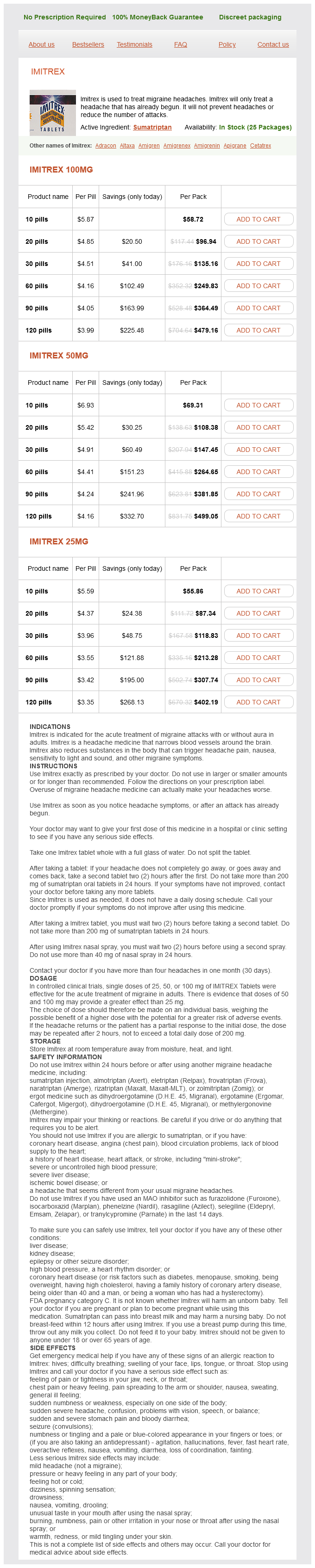
Migraines are a typical type of headache that impacts millions of individuals worldwide. These headaches may be debilitating, causing severe pain, nausea, and sensitivity to gentle and sound. For those who undergo from frequent migraines, discovering an acceptable therapy is often a difficult and irritating experience. However, there's a medication that has been confirmed to be effective in treating migraines - Sumatriptan, generally sold under the brand name Imitrex.
Sumatriptan is obtainable in a number of forms, together with tablets, nasal sprays, and injections. The tablets are the most popular form and are available available in the market in varied strengths. For delicate to reasonable migraine attacks, a lower dose of 25mg or 50mg is beneficial, whereas the next dose of 100mg is appropriate for extra severe assaults. The nasal spray and injections are really helpful for those who experience nausea and vomiting throughout a migraine attack, as they are faster and more effective in providing reduction.
Discontinuous capillaries (sinusoidal capillaries spasms after urinating buy sumatriptan 25 mg mastercard, sinusoids) have large openings in their endothelial cells and are separated by wide irregular intercellular gaps. Also, endothelial cells rest on a discontinuous basal lamina, which in some organs is rudimental and may be absent. These observations support the suggested mode of fenestration formation described above. Discontinuous capillaries (also called sinusoidal capillaries or sinusoids) are typically found in the liver, spleen, and bone marrow. They are larger in diameter and more irregularly pericyte shaped than other capillaries. Structural features of these capillaries vary from organ to organ and include specialized cells. Kupffer cells (stellate sinusoidal macrophages) and vitamin Astoring Ito cells (hepatic stellate cells) in the liver occur in association with the endothelial cells of hepatic sinuses. In the spleen, endothelial cells exhibit a unique spindle shape with gaps between the neighboring cells; the basal lamina underlying the endothelium is rudimental and may be partially or even completely absent. Pericytes represent a population of undifferentiated mesenchymal stem cells that are associated with capillaries. The endothelial cells that make up the wall of a continuous capillary contain numerous pinocytotic vesicles. The cell junctions are frequently marked by cytoplasmic (marginal) folds that protrude into the lumen. The endothelial cell nuclei are not included within the plane of section in the micrograph. Similarly, the electron micrograph shows only a small amount of pericyte cytoplasm. Capillaries and some postcapillary venules are associated with perivascular cells exhibiting cellular processes that wrap around vascular endothelial cells. They intimately surround the capillary, with branching cytoplasmic processes, and are enclosed by a basal lamina that is continuous with that of the endothelium. There is some evidence to suggest that pericytes can modulate capillary blood flow in specific capillary beds. Pericytes provide vascular support and promote stability of capillaries and postcapillary venules through complex, bidirectional physical and chemical communication with vascular endothelial cells. Histologically, pericytes display features of undifferentiated mesenchymal stem cells with large nuclei rich in heterochromatin. Experiments have shown that environmental signals can stimulate proliferation, migratory capability, and differentiation of pericytes into a variety of cell types, including adipocytes, fibroblasts, chondrocytes, osteocytes, and skeletal muscle cells. In this condition, capillary pressure can decrease and greatly increase absorption of tissue fluid. This situation occurs during loss of blood volume and can add considerable amount of fluid into the blood, preventing hypovolemic shock. The density of the capillary network determines the total surface area available for exchange between the blood and tissue. The liver, kidney, cardiac muscle, and skeletal muscle have rich capillary networks. Dense connective tissue is less metabolically active and has less extensive capillary networks. The cytoplasm of the endothelial cells contains numerous fenestrations (small arrows). In some of the thicker regions of the endothelial cells where the fenestrations are absent, pinocytotic vesicles are present. Part of a pericyte is seen on the bottom of the electron micrograph, including its nucleus in the lower left corner of the micrograph. The inset shows to advantage the fenestrations and the diaphragm that spans the openings (large arrows). Pericytes are directly involved in the pathogenesis of vascular driven diseases. In addition, uncontrolled divisions of pericytes give rise to the hemangiopericytoma, a rare vascular tumor that can originate in the body anywhere there are capillaries. Generally, in a microvascular bed, arteries convey blood to the capillaries, and veins convey blood away from the capillaries. However, all the blood does not necessarily pass from arteries to capillaries and then to veins. In many tissues, there are direct routes between the arteries and veins that divert blood from the capillaries. Although capillaries themselves have no smooth muscle in their walls, a sphincter of smooth muscle called the precapillary sphincter is located at their origin from either an arteriole or a metarteriole. Pressure within the capillaries increases, and much of the plasma fluid is driven into the tissue. Small veins are less than 1 mm in diameter and are con- tinuous with muscular venules. Pericyte coverage is more extensive in the postcapillary venules than in the capillaries. High endothelial venules are specialized postcapillary venules found in lymphoid tissues that support high levels of lymphocyte migration from the blood.
Glucagon stimulates release of glucose into the bloodstream and stimulates gluconeogenesis (synthesis of glucose from metabolites of amino acids) and glycogenolysis (breakdown of glycogen) in the liver muscle relaxant and painkiller purchase sumatriptan visa. Glucagon also stimulates proteolysis to promote gluconeogenesis, mobilizes fats from adipose cells (lipolysis), and stimulates hepatic lipase. Blood glucose levels below 70 mg/100 mL stimulate release of glucagon; blood glucose levels significantly above 70 mg/100 mL inhibit glucagon secretion. Insulin inhibits release of glucagon by A cells, but because of the cascading circulation in the islet (explained in a following paragraph), this inhibition is affected by a hormonal action of insulin carried in the general circulation. About 10% of the islet cells have nerve endings directly on their plasma membrane. Ionic events triggered by synaptic transmitters at the nerve endings are carried from cell to cell across these junctions. It is identical to the hormone secreted by the hypothalamus that regulates somatotropin (growth hormone) release from the anterior pituitary gland. Although the precise role of somatostatin in the islets is unclear, it has been shown to inhibit both insulin and glucagon secretion. The molecular characteristics of the major and some minor islet hormones are summarized in Table 18. Some amino acids also stimulate insulin secretion, either alone or in concert with elevated blood glucose levels. This neural control of insulin and glucagon may contribute to the availability of circulating glucose in stress reactions. The blood supply to the pancreas provides a cascading perfusion of the islets and acini. As mentioned earlier, islets of Langerhans comprise only 1% to 2% of the total mass of the pancreas; however, they receive about 10% to 15% of the pancreatic blood flow. The most common pattern is when blood enters the islets at the center, initially perfusing the core of the islet and then spreading to the periphery. In the second pattern, several arterioles enter the periphery of the islets and branch into fenestrated capillaries to perfuse the core of the islet. In humans, it is likely that the capillaries first perfuse the A and D cells, peripherally, before the blood reaches the B cells, centrally. Larger vessels that travel in septa that penetrate the central portion of the islet are also accompanied by A and D cells, so that blood reaching the B cells has always first perfused the A and D cells. Recent in vivo fluorescence imaging studies show distinct blood flow dynamics in the islets of Langerhans. These studies suggest that blood flow is regulated by the blood glucose levels in addition to the complex interaction among vasodilators and vasoconstrictors, gastrointestinal peptides, and the autonomic nervous system. Large efferent capillaries leave the islet and branch into the capillary networks that surround the acini of the exocrine pancreas. This cascading flow resembles the portal systems of other endocrine glands (pituitary, adrenal). It is a small protein consisting of two polypeptide chains joined by disulfide bridges. Its biosynthesis presents a clear example of the importance of posttranslational processing in the achievement of the final, active structure of a protein. Insulin is originally synthesized as a single 110amino acid polypeptide chain with a molecular weight of about 12,000 Da. Posttranslational processing reduces the preproinsulin to a polypeptide with a molecular weight of about 9,000 Da. It is stored in the secretory vesicles and released with the insulin in equimolar amounts. Because C peptide has a longer half-life than insulin, higher concentrations of C peptides are detected in the peripheral blood. For these reasons, measurement of circulating levels of C peptides provides important clinical information about the secretory activity of B cells. Because C peptide is cleared from the body by the kidney, measurement of its urinary excretion provides useful information about B-cell insulin secretion. C peptide measurements are frequently used to assess the residual B-cell function in patients treated with insulin, to distinguish between types 1 and 2 diabetes, and in the diagnosis and monitoring therapy of insulinoma (tumor of B cells). C peptide may also be used to monitor the progress of pancreas or islet cell transplantation. Insulin is synthesized as the preproinsulin, a single polypeptide chain that undergoes posttranslational modifications. The liver plays an important role in the uptake, storage, and distribution of nutrients. The liver also acts as an exocrine organ (produces bile) and performs endocrine-like functions. The liver has a dual blood supply: a venous supply via the hepatic portal vein and an arterial supply via the hepatic artery. Hepatocytes (as seen in the classic lobule) are organized into irregular anastomosing plates that radiate toward a central vein. Corners of the polygonal classic lobule are occupied by the portal triad, which contains branches of portal vein, hepatic artery, bile ducts, and small lymphatic vessels. Hepatic sinusoids form irregular vascular channels that run parallel and between the plates of hepatocytes. They receive mixed blood (75%) from venous portal circulation and arterial blood (25%) from systemic circulation. The perisinusoidal space (space of Disse) lies between hepatocytes and the endothelium; it is the site of exchange of materials between blood and liver cells. The sinusoidal endothelium includes specialized stellate sinusoidal macrophages (Kupffer cells), which remove senile red blood cells and recycle iron molecules.
Sumatriptan Dosage and Price
Imitrex 100mg
- 10 pills - $58.72
- 20 pills - $96.94
- 30 pills - $135.16
- 60 pills - $249.83
- 90 pills - $364.49
- 120 pills - $479.16
Imitrex 50mg
- 10 pills - $69.31
- 20 pills - $108.38
- 30 pills - $147.45
- 60 pills - $264.65
- 90 pills - $381.85
- 120 pills - $499.05
Imitrex 25mg
- 10 pills - $55.86
- 20 pills - $87.34
- 30 pills - $118.83
- 60 pills - $213.28
- 90 pills - $307.74
- 120 pills - $402.19
On the basis of their appearance after staining muscle relaxant use in elderly cheap sumatriptan online master card, leukocytes are traditionally divided into granulocytes (neutrophils, eosinophils, and basophils) and agranulocytes (lymphocytes and monocytes). Although both cell types may contain granules, the granulocytes possess obvious, specifically stained granules in their cytoplasm. Scientists originally thought that the fine neutrophil granules were stained by a "neutral dye" that formed when methylene blue and its related azures were combined with eosin. The mechanism by which the specific neutrophil granules are stained is still not clearly understood. Some of the basic dyes (the azures) are metachromatic and may impart a violet to red color to the material they stain. The shape of the erythrocyte is maintained by a specialized cytoskeleton that provides the mechanical stability and flexibility necessary to withstand forces experienced during circulation. As erythrocytes in circulation navigate through a small network of capillaries, they are exposed to high amounts of shear force that cause them to undergo rapid and reversible deformations. To cope with this stress, the erythrocyte cell membrane has a unique cytoskeletal structure. In addition to a typical lipid bilayer, it contains two functionally significant groups of proteins. They function only within the bloodstream to bind oxygen for delivery to the tissues and, in exchange, bind carbon dioxide for removal from the tissues. In a healthy individual, approximately 1% of erythrocytes are removed from circulation each day due to senescence (aging); however, bone marrow continuously produces new erythrocytes to replace those lost. The majority of aged erythrocytes (90%) are phagocytosed by macrophages in the spleen, bone marrow, and liver. The remaining aged erythrocytes (10%) break down intravascularly, releasing insignificant amounts of hemoglobin into the blood. The extracellular domains of these integral membrane proteins are glycosylated and express specific blood group antigens. Glycophorin C, a member of the glycophorin family of transmembrane proteins, plays an important role in attaching the underlying cytoskeletal protein network to the cell membrane. Band 3 protein is the most abundant transmembrane protein in erythrocyte cell membrane. They are organized into a two-dimensional hexagonal lattice network that laminates the inner layer of the membrane. The lattice itself, which is positioned parallel to the membrane, is composed mainly of cytoskeletal proteins, including -spectrin and -spectrin molecules. They assemble to form an antiparallel heterodimer held together by multiple lateral bonds. Dimers then associate in a head-to-head formation to produce long and flexible tetramers. The spectrin filaments are anchored to the lipid bilayer by two large protein complexes. The erythrocyte is an anucleated cell in a shape of a biconcave disc containing hemoglobin. The surface area of an erythrocyte is about 140 m2 and its mean corpuscular (cell) volume ranges from 80 to 99 fL (1 fL 10 15 L). This unique cytoskeletal arrangement contributes to the shape of the erythrocyte and imparts elastic properties and stability to the membrane. Photomicrograph of three capillaries (Cap) joining to form a venule (V), as observed in adipose tissue within a full-thickness mesentery spread. The erythrocytes appear in single file in one of the capillaries (the other two are empty). The light center area of some of the erythrocytes results from their biconcave shape. Erythrocytes are highly plastic and can fold on themselves when passing through very narrow capillaries. The stacks of erythrocytes in these preparations are not unusual and are referred to as rouleau. The rectangle in the sectioned erythrocyte (upper right) represents the area of membrane in the larger diagram. The large diagram shows the arrangement of peripheral and integral membrane proteins. The integral membrane protein glycophorin C associates with peripheral membrane band 4. These peripheral complexes interact with spectrin to form a cytoskeletal hexagonal lattice immediately adjacent to the cytoplasmic surface of the plasma membrane. Spectrin lattice with peripheral membrane protein complexes is anchored to the plasma membrane by the glycophorin C and band 3 proteins, which, on the extracellular surface, are glycosylated and support the majority of carbohydrate-defined blood group antigens. Any defect in the expression of genes that encode these cytoskeleton proteins can result in abnormally shaped and fragile erythrocytes. For example, hereditary spherocytosis is caused by an autosomal dominant mutation of proteins that function in anchoring erythrocyte plasma membrane to the cytoplasm. In this condition, erythrocyte plasma membrane has defective anchor points, causing it to detach and peel off from the cytoplasm. Another erythrocyte membrane abnormality, hereditary elliptocytosis, is caused by one of several autosomal dominant mutations affecting spectrin molecules. Plasma membrane in affected cells fails to rebound from deformations and progressively elongates, resulting in the formation of elliptical erythrocytes. In both conditions, erythrocytes are unable to adapt to changes in their environment. Erythrocytes contain hemoglobin, a protein specialized for the transport of oxygen and carbon dioxide.

ADDII BIOTECH is proud to announce that the company's manufacturing facility located at Baddii, Himachal Pradesh has recently been approved by Food & Drug Authority of Ghana.
Our Manufacturing capability includes a wide range of therapeutic products covering almost every segment.
ADDII BIOTECH given our strong emphasis on product quality and services.