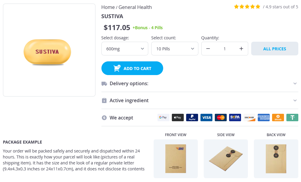
Sustiva
General Information about Sustiva
Despite its effectiveness, Sustiva is not without limitations. It could interact with different drugs, including over-the-counter supplements, and might cause birth defects if taken throughout being pregnant. Therefore, it is essential to tell a healthcare provider of all medications being taken before beginning Sustiva.
In conclusion, Sustiva is a priceless treatment option for people residing with HIV-1. Its accessibility, once-daily dosing, and effectiveness in suppressing the virus make it an important component of ART. As analysis and development within the area of HIV treatment continue, we are able to hope to see extra developments like Sustiva in the battle in opposition to this global well being problem.
One of the benefits of Sustiva is its lengthy half-life, which means that it remains active within the body for an extended time period. This permits for once-daily dosing, making it easier for sufferers to adhere to their remedy routine. Adherence to a therapy plan is essential for the success of ART and to forestall the development of drug resistance.
When utilized in combination with other antiretroviral medications, Sustiva has been proven to successfully suppress HIV and enhance the number of CD4 T-cells within the body. It has additionally been related to a lower in HIV-related diseases and deaths.
Like any treatment, Sustiva might cause side effects. Common unwanted effects embody dizziness, bother sleeping, drowsiness, and vivid goals. These unwanted effects are usually delicate and tend to enhance with continued use. However, in uncommon instances, more extreme unwanted aspect effects could happen, corresponding to severe skin reactions, liver problems, and psychiatric symptoms. It is important to report any unusual side effects to a healthcare supplier.
Sustiva, also called efavirenz, is an antiviral agent and a non-nucleoside reverse transcriptase inhibitor (NNRTI) used in the therapy of human immunodeficiency virus sort 1 (HIV-1). It was first approved by the United States Food and Drug Administration (FDA) in 1998 and is manufactured by Bristol-Myers Squibb in collaboration with United Drug.
A important concern when treating HIV is the potential for drug resistance. This occurs when the virus mutates and turns into immune to the consequences of the medicine. To cut back the danger of drug resistance, Sustiva is usually mixed with different antiretrovirals to create a potent and efficient therapy routine.
Sustiva is available in 200 mg capsules and is usually taken once a day on an empty stomach. It is usually prescribed as a part of a mix remedy for HIV, together with other antiretroviral medicines. This is named antiretroviral remedy (ART) and is important for managing HIV and stopping development to AIDS.
HIV-1 is a virus that attacks the immune system, specifically the CD4 T-cells that are liable for combating infection. Without treatment, HIV can progress to acquired immune deficiency syndrome (AIDS), which is a life-threatening situation. Sustiva works by preventing the virus from multiplying and thus decreasing the quantity of HIV within the physique.
The gastric antrum extends from its indistinct border with the body to the junction of the pylorus with the duodenum medicine 834 order sustiva mastercard. These gross anatomic landmarks correspond roughly with the mucosal histology because antral mucosa (pyloric gland mucosa) actually extends from an area on the lesser curvature somewhat above the incisura. The pylorus (pyloric channel) is a tubular structure joining the duodenum to the stomach and contains the palpable circular muscle, namely the pyloric sphincter. The pylorus is somewhat mobile owing to its enclosure between the peritoneum of the greater and lesser omenta but is generally located 2 cm to the right of midline at L1. Corresponding motor and secretory functions of these regions of the stomach are discussed in detail in Chapters 50 and 51. From these plexuses, postganglionic fibers are distributed to cells and glands and to smooth muscle. Four layers make up the gastric wall: mucosa, submucosa, muscularis propria, and serosa. Mucosa lines the gastric lumen, appearing as a smooth, velvety, blood-filled lining. The mucosa of the cardia, antrum, and pylorus is somewhat paler than that of the fundus and body. It is within the fundic and body mucosa that most of the functional secretory elements of the stomach are located (see Chapter 51). The submucosa, immediately deep to the mucosa, provides the dense connective tissue skeleton of collagen and elastin fibers. Lymphocytes, plasma cells, arterioles, venules, lymphatics, and the submucosal plexus are also contained within the submucosa. The third tissue layer, the muscularis propria, is a combination of 3 muscle layers: inner oblique, middle circular, and outer longitudinal. The inner oblique muscle fibers course over the gastric fundus, covering the anterior and posterior aspects of the stomach wall; the middle circular fibers encircle the body of the stomach, thickening distally to become the pyloric sphincter; and the outer longitudinal muscle fibers course primarily along the greater and lesser curvatures of the stomach. The final layer of the stomach is the transparent serosa, a continuation of the visceral peritoneum. The cells secrete mucus in granules that are released via exocytosis, apical expulsion, and cell exfoliation. The primary role of mucus, along with bicarbonate, is luminal cytoprotection from "the elements": acid, pepsin, ingested substances, and pathogens. The surface epithelial lining is invaginated by gastric pits, or foveolae, that provide the gastric glands access to the gastric lumen, with a ratio of 1 pit to 4 or 5 gastric glands. The first region, the cardia, is a small transition zone from esophageal squamous epithelium to gastric columnar epithelium. The cardia has been a controversial histologic area of discussion, with theories suggesting that its presence is pathologic. However, recent observations concluded that cardiac mucosa develops during gestation and is present at birth. There is a gradual transition from cardiac glands to the second region, the acid-secreting segment of the stomach. This region encompasses the gastric fundus and body and contains the parietal (or oxyntic or fundic) glands. Parietal, chief (also known as peptic), endocrine, mucous neck, and undifferentiated cells compose the oxyntic glands. The final region, corresponding to the antrum and pylorus, contains the pyloric glands, composed of endocrine cells, including gastrinproducing G cells and mucous cells. These fairly straight and simple tubular glands are closely associated in the areas of gastric fundus and body. A typical gland is subdivided into 3 areas: the isthmus (where surface mucous cells predominate), the neck (where parietal and mucous neck cells predominate), and the base (where chief cells predominate, along with some parietal and mucous neck cells). Parietal cells bulge into the lumina of the oxyntic glands and, as the primary hydrogen secretors, have ultrastructural characteristics different from other gastric cells: large mitochondria, microvilli lacking in glycocalyx, and a cytoplasmic canaliculi system in contact with the lumen. In the nonsecreting parietal cell, a cytoplasmic tubulovesicular system predominates and short microvilli line the apical canaliculus. In the secreting state, the tubulovesicular system disappears, leaving an extensive system of intracellular canaliculi containing long microvilli. Additionally, parietal cells are the site of intrinsic factor secretion via membrane-associated vesicle transport. Closely associated with parietal cells are mucous neck cells, which appear singly close to parietal cells or in groups of 2 or 3 in the oxyntic gland neck or isthmus. Mucous neck cells differ from their surface counterparts in their synthesis of acidic, sulfated mucus rather than the neutral mucus. Additionally, mucous neck cells have basal nuclei and larger mucous granules around the nucleus, rather than apically located granules. Function of the 2 cell types appears different in that surface mucous cells are cytoprotective, whereas the mucous neck cell functions as a stem cell precursor for surface mucous, parietal, chief, and endocrine cells. Chief cells, also known as zymogen cells, predominate in deeper layers of the oxyntic glands. The cytoplasm of chief cells has prominent basophilic staining owing to abundant ribosomes; these ribosomes are either free in the cytoplasm or in association with an extensive endoplasmic reticulum system. Zymogen granules lie in the apical cytoplasm; their contents are released into the gastric lumen following fusion of the limiting membrane of the granule with the luminal membrane. A variety of endocrine, or enteroendocrine, cells are scattered among the cells of the oxyntic glands (see Chapter 4). These cells vary in location, being either open or closed relative to the gastric lumen.
Faecal microbiota profiles as diagnostic biomarkers in primary sclerosing cholangitis symptoms dehydration buy sustiva online from canada. The gut microbial profile in patients with primary sclerosing cholangitis is distinct from patients with ulcerative colitis without biliary disease and healthy controls. Characterisation of the faecal microbiota in Japanese patients with paediatric-onset primary sclerosing cholangitis. Distinct gut microbiota profiles in patients with primary sclerosing cholangitis and ulcerative colitis. Predictors of pouchitis after ileal pouch-anal anastomosis: a retrospective review. Inflammatory bowel disease is associated with poor outcomes of patients with primary sclerosing cholangitis. Primary sclerosing cholangitis: natural history, prognostic factors and survival analysis. Natural history and prognostic factors in 305 Swedish patients with primary sclerosing cholangitis. Primary sclerosing cholangitis: clinical presentation, natural history and prognostic variables: an Italian multicentre study. Primary sclerosing cholangitis associated with inflammatory bowel disease in Cape Town, 1975 1981. Prevalence of sclerosing cholangitis detected by magnetic resonance cholangiography in patients with long-term inflammatory bowel disease. The natural history of primary sclerosing cholangitis in 781 children: a multicenter, international collaboration. Primary sclerosing cholangitis, autoimmune hepatitis, and overlap in Utah children: epidemiology and natural history. A 2-year follow-up study of anti-neutrophil antibody in primary sclerosing cholangitis: relationship to clinical activity, liver biochemistry and ursodeoxycholic acid treatment. Antineutrophil antibodies define clinical and genetic subgroups in primary sclerosing cholangitis. Radiologic course of primary sclerosing cholangitis: assessment by three-dimensional magnetic resonance cholangiography and predictive features of progression. Low prevalence of alterations in the pancreatic duct system in patients with primary sclerosing cholangitis. Clinical relevance of perihepatic lymphadenopathy in acute and chronic liver disease. Baseline values and changes in liver stiffness measured by transient elastography are associated with severity of fibrosis and outcomes of patients with primary sclerosing cholangitis. Comparison of the clinicopathologic features of primary sclerosing cholangitis and primary biliary cirrhosis. Morphologic features of chronic hepatitis associated with primary sclerosing cholangitis and chronic ulcerative colitis. A preliminary trial of highdose ursodeoxycholic acid in primary sclerosing cholangitis. Efficacy and safety of simtuzumab for the treatment of primary sclerosing cholangitis: results of a phase 2b, dose-ranging, randomized, placebo-controlled trial. Validation of the prognostic value of histologic scoring systems in primary sclerosing cholangitis: an international cohort study. Applicability and prognostic value of histologic scoring systems in primary sclerosing cholangitis. Application of a new histological staging and grading system for primary biliary cirrhosis to liver biopsy specimens: interobserver agreement. Factors that reduce health-related quality of life in patients with primary sclerosing cholangitis. Pruritus is associated with severely impaired quality of life in patients with primary sclerosing cholangitis. Factors that influence health-related quality of life in patients with primary sclerosing cholangitis. Oral naltrexone treatment for cholestatic pruritus: a double-blind, placebo-controlled study. Serum autotaxin is increased in pruritus of cholestasis, but not of other origin, and responds to therapeutic interventions. The impact of fragility fractures on health-related quality of life in patients with primary sclerosing cholangitis. Influence of dominant bile duct stenoses and biliary infections on outcome in primary sclerosing cholangitis. Risk factors and clinical presentation of hepatobiliary carcinoma in patients with primary sclerosing cholangitis: a case-control study. Cholangiocarcinoma in patients with primary sclerosing cholangitis: a multicenter case-control study. Sensitivity of endoscopic retrograde cholangiopancreatography standard cytology: 10-yr review of the literature. A prospective, randomized-controlled pilot study of ursodeoxycholic acid combined with mycophenolate mofetil in the treatment of primary sclerosing cholangitis. Ursodeoxycholic acid therapy for primary sclerosing cholangitis: results of a 2-year randomized controlled trial to evaluate single versus multiple daily doses. Metronidazole and ursodeoxycholic acid for primary sclerosing cholangitis: a randomized placebo-controlled trial. High dose ursodeoxycholic acid for the treatment of primary sclerosing cholangitis is safe and effective. A double-blind, placebo-controlled, randomized study of infliximab in primary sclerosing cholangitis. No superiority of stents vs balloon dilatation for dominant strictures in patients with primary sclerosing cholangitis.
Sustiva Dosage and Price
Sustiva 600mg
- 10 pills - $130.06
- 20 pills - $252.05
- 30 pills - $357.03
- 60 pills - $648.03
Sustiva 200mg
- 30 pills - $158.07
- 60 pills - $266.07
- 90 pills - $361.06
- 120 pills - $475.04
D symptoms xanax buy generic sustiva 200 mg, An intraoperative picture of the gastric duplication after dissection of the stomach and before resection. Gastric Teratoma Gastric teratomas are benign neoplasms of the stomach that occur almost exclusively in males. These tumors may have their origins in pluripotential cells and contain all 3 embryonic germ cell layers. Most are located along the greater curvature of the stomach and are extragastric, although intramural extension has been reported. In virtually all cases, gastric teratoma is an isolated finding and is not associated with other tumors or malformations. Premalignant changes and frank malignant transformation to adenocarcinoma have been reported,30,31 and peritoneal gliomatosis has been observed. Fortunately, even those cases with malignant histologic features or extension into adjacent tissues have an excellent prognosis. The newborn infant with a teratoma may be delivered prematurely or have respiratory distress on the basis of increased abdominal pressure. Delivery may be difficult, putting the infant at risk for injuries such as shoulder dystocia. Gastric teratoma associated with gastric perforation, mimicking meconium peritonitis, has Gastric Volvulus See Table 49. A localized lack of nitric oxide synthase, an enzyme associated with smooth muscle relaxation, or abnormal neuronal innervation associated with decreased muscle neurofilaments, nerve terminals, synaptic vesicle protein, and neural cell adhesion molecule35 has been implicated; however, anatomic studies cannot determine whether nitric oxide synthase deficiency is a primary or secondary event,36 and nitric oxide synthase deficiency is only notable in a subset of cases. Incidence is highest among whites (especially northern Europeans), whereas incidence is lower among African Americans and Africans and lowest among Asians. Others at increased risk are first-born male infants, especially those with high birth weights or born to professional parents. Initially, infants present with mild spitting, which progresses to projectile vomiting following feedings. Vomiting may be so forceful as to exit through the nostrils, as well as the mouth. Emesis may contain "coffee ground" material or small amounts of frank blood, but is rarely bilious. Early in the course, the infant remains hungry following vomiting episodes but, with time, loses interest in feeding and may present wasted and severely volume depleted. The classic physical signs are a palpable pyloric mass and visible peristaltic waves. The palpable "olive" is most easily felt in a wasted patient, immediately following emesis or aspiration of the stomach. The location of the olive varies from the level of the umbilicus to near the epigastrium. The pyloric mass is palpable in 70% to 90% of affected infants, depending on the experience and patience of the examiner. Emptying the stomach by nasogastric tube placement and palpation of the stomach with the infant in the prone position may enhance detection. Many infants appear jaundiced due to an indirect hyperbilirubinemia related to volume depletion and, perhaps, malnutrition. When the presentation is typical and the olive palpated, no studies are necessary. However, in the minority of infants with projectile vomiting, definitive diagnosis requires radiologic studies. Non-contrast radiography demonstrates a distended stomach with paucity of gas beyond the stomach. The numeric value for the lower limit of pyloric muscle thickness has varied in reports in the literature, ranging between 3 and 4. Many consider the numeric value less important than the overall morphology of the canal and real-time observations. Contrast radiography must be performed carefully, and gastric contents should first be aspirated. Characteristic findings include an elongated narrow pylorus with the appearance of a "double channel. In a few cases, no etiology is determined; it is therefore unknown whether these are missed infantile cases or whether the hypertrophy occurred later in life. The resected pylorus demonstrates normal mucosa and marked circumferential thickening of the muscularis propria. In contrast with the infantile form, the physical examination may not be helpful because the pyloric mass is difficult to palpate in adults. On contrast radiography, the elongated narrow pylorus is again apparent; gastric emptying is delayed, and the stomach may be dilated. Treatment Traditionally, surgical pyloromyotomy or resection of the involved region has been considered the procedure of choice. Because of the risk of a small focus of carcinoma, surgical resection of the pylorus has been recommended. Depending on severity, fluid and electrolyte repletion can usually be accomplished within 24 hours. Definitive therapy is the Ramstedt pyloromyotomy, which entails a longitudinal incision through the hypertrophied pyloric muscle down to the submucosa on the anterior surface of the pylorus. After spreading the muscle, the intact mucosa bulges through the incision to the level of the incised muscle.


