
Terbinafine
General Information about Terbinafine
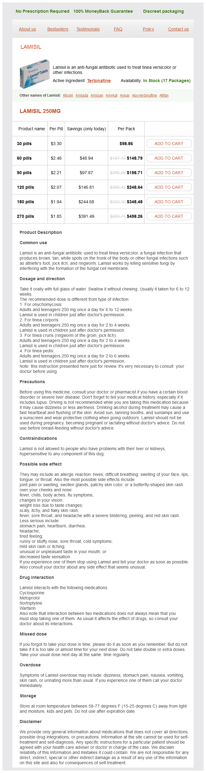
Terbinafine, marketed beneath the brand name Lamisil, is an anti-fungal antibiotic that is generally used to deal with fungal infections. It is primarily used to deal with tinea versicolor, a standard skin infection brought on by yeast, however may also be effective against different forms of fungal infections.
It is important to notice that while terbinafine is effective in opposition to fungal infections, it doesn't have any impact on bacterial or viral infections. Therefore, it shouldn't be used to treat circumstances corresponding to colds or the flu. It can be not beneficial to be used in pregnant or breastfeeding ladies, as the effects on the unborn child are not totally understood.
Lamisil is out there as both an oral medication and a topical cream. The oral type is usually prescribed for tinea versicolor, as it can successfully attain the deeper layers of the pores and skin where the fungus may be present. It is taken once a day for a period of two to four weeks, relying on the severity of the an infection. The topical cream is often prescribed for milder cases or as a maintenance therapy to forestall future outbreaks. It is applied on to the affected areas once or twice a day, relying on the instructions supplied by the doctor.
In conclusion, terbinafine is an effective medication for treating fungal infections, notably tinea versicolor. With its capability to inhibit the growth of fungi and clear up pores and skin infections, it has become a well-liked alternative for both docs and patients. However, it's important to use this medicine as directed by a healthcare skilled and to be aware of any potential side effects. If you're experiencing symptoms of a fungal an infection, seek the advice of together with your doctor to discover out if terbinafine could be the proper remedy choice for you.
In addition to treating tinea versicolor, terbinafine has also been found to be effective in treating different forms of fungal infections such as athlete's foot, jock itch, and ringworm. These infections occur when fungi invade the highest layer of the skin, inflicting it to become purple, itchy, and inflamed. By destroying the fungal cells, terbinafine might help to clear up these infections and supply reduction from uncomfortable symptoms.
Terbinafine works by inhibiting the expansion of the fungus liable for the an infection. It does this by interfering with the manufacturing of ergosterol, an essential component of fungal cell membranes. Without ergosterol, the fungal cells are weakened and eventually die, permitting the an infection to clear up.
Terbinafine is usually well-tolerated, and most of the people experience few unwanted side effects. The commonest side effects which were reported embody rash, itching, and gastrointestinal issues corresponding to nausea and diarrhea. Rarely, extra critical side effects such as liver harm have been reported, so it is essential to comply with the prescribed dosage and monitor any adjustments in your health whereas taking terbinafine.
Tinea versicolor, also called pityriasis versicolor, is a skin infection that's attributable to the yeast Malassezia furfur. This fungus is naturally found on healthy skin, however in some cases, it could possibly overgrow and trigger discolored patches on the skin. These patches may be lighter or darker than the encompassing pores and skin and could additionally be accompanied by delicate itching. Tinea versicolor is more common in warm and humid climates and can have an result on people of any age, however is mostly seen in adolescents and young adults.
Transcranial cerebral oximetry is a noninvasive technique for monitoring changes in cerebral oxygenation antifungal otc cream buy terbinafine with a visa. Changes in transcranial cerebral oximetry result from changes in the balance between the cerebral oxygen supply and oxygen consumption. The metabolic state of the brain can also be assessed with cerebral microdialysis, which monitors energy metabolites over extended periods, but only in a small focal volume of the brain. The threshold for abnormal metabolism is evidenced by the lactate/pyruvate ratio of 40. One conundrum that remains unsolved, even with multimodality monitoring, is the value of continuous information versus global information about the condition of the brain. Autoregulation and compliance are two other measures that help us to individualize management and set different thresholds for each patient. Hypoxia from adult respiratory distress syndrome or ventilator-associated pneumonia and hyperthermia from infection or sepsis are the most common. Comatose patients are at high risk for deep vein thrombosis and pulmonary embolism. After the first 48 hours subcutaneous heparin or lowmolecular-weight heparin can be used safely. To achieve and maintain normothermia, advanced cooling methods, surface cooling, or intravascular cooling needs to be used. Conventional cooling methods, ice packs, and Tylenol have not shown to be successful (Badjatia et al. While methods such as intravascular cooling and surface cooling have shown to be effective for lowering body temperature, intravascular cooling is more effective in keeping the temperature in a normothermic range (Hoedemaekers et al. However, the development of mass effect may result in secondary brain injury, posing the risk of further neurological deterioration, herniation, and death. Various surgical techniques have been advocated to evacuate intracranial hemorrhages. The most commonly used is craniotomy, with or without subtemporal decompressive craniectomy. Sometimes a large decompressive hemicraniectomy is performed at the same time if the brain appears swollen intraoperatively. More midline shift than one would expect from the extra-axial hematoma, effacement of basal cisterns and sulci, and loss of graywhite matter differentiation are warning signs. This is generally followed by focal neurological signs such as hemiparesis, seizures, pupillary dilation, decrease in consciousness, and, in the worst-case scenario, decortication. The following Craniocerebral Trauma 879 guidelines list the criteria for surgical evacuation of an acute epidural hematoma (Bullock et al. The guidelines for surgical management of depressed skull fractures recommend administering antibiotics routinely. Most common are deficiencies in growth hormone, gonadotropin, and corticotropin levels. Post-traumatic communicating hydrocephalus has an incidence of 14% in severe head injuries. Approaches used to treat neurocognitve deficits have been cognitive and behavioral therapy with pharmacological treatment. Psychosocial problems are a persistent long-term problem and can interfere with rehabilitation. This situation is aggravated by a lack of opportunity to establish new social contacts and friends. Clinicians such as psychiatric social workers, psychologists, or psychiatrists may need to be called upon more often to provide the psychological services that may be necessary for many of these patients. Neurological deterioration over time is common and also can influence the decision to operate. The magnitude of preoperative midline shift also seems to correlate with unfavorable outcome (Nelson et al. Isolated linear fractures are usually of little clinical significance unless they are near or traverse a suture or they involve a venous sinus groove or vascular channel. Although rare, a growing linear skull fracture can develop in young children, especially if the parietal bone is fractured. Patients with simple ("closed") depressed cranial fractures may be treated nonoperatively if there is no clinical or radiographic evidence of dural penetration (pneumocephalus), significant intracranial hematoma, depression >1 cm, frontal sinus involvement, cosmetic deformity, or gross wound contamination. Compound (open) depressed skull fractures are associated with infections and the development of late epilepsy. They undergo surgical debridement and elevation to decrease the possibility of infection. Depending on the statistical analysis performed, one study found worse outcome above 39 and 65 years of age (Kuhne et al. Multiple regression analysis showed that above this threshold every 10-year increase in age leads to a 10% increase in mortality. Even with timely and satisfactory surgery, unexplained clinical deterioration occurs in patients 70 years or older. Additionally, falls can cause other injuries such as hip fractures, which can impede independent living and increase the risk of premature death. However, rehabilitation efforts in the elderly have been poor because of the often-negative attitude (ageism) toward the recovery potential in this group. Despite their relative disadvantages, the elderly might see improved outcomes with rehabilitation programs intended for their age group. They have been externally validated and demonstrated good generalizability (Roozenbeek et al. How and if these models are accurate enough to be applied to clinical practice may be decided by the clinicians.
After gadolinium administration (E) antifungal eo purchase 250 mg terbinafine otc, extensive enhancement within the area of infarct indicates breakdown of the bloodbrain barrier. As infarction develops, however, other symptoms such as seizures develop, leading to hemorrhagic conversion of the infarction, herniation, and death. Although still controversial, most studies suggest that anticoagulation of the patient with sagittal sinus thrombosis is indicated even when there is hemorrhage into the brain. This edema is both cytotoxic (direct damage to cells) and vasogenic (inflammatory response induced by toxic blood products). Growth of hematoma was observed after 24 hours in 38% of patients who were imaged within 3 hours of hemorrhage onset and again within 24 hours (Brott et al. Secondary hemorrhagic transformation can occur in an area of infarction, particularly when the ischemic region is large. Generally, the hemorrhagic transformation is found 24 to 72 hours after the insult. Accumulation of blood causes both mass effect on the surrounding tissues and release of toxic blood products into adjacent tissues. Blood contains coagulation cascade enzymes such as thrombin and plasmin, which are pluripotential molecules that can damage cells both directly by their toxic effects and indirectly by activation of other proteases. In experimental animals, injection of thrombin into the brain produces a focal increase in brain water content (Xi et al. A, T2-weighted magnetic resonance imaging shows extensive cerebral edema in posterior white matter regions, with less involvement of the gray matter. B, A higher level of the same scan sequence as in A, showing some frontal lobe involvement. During treatment of the diabetic ketoacidosis, blood osmolality is reduced, and water moves into brain along the osmotic gradient, resulting in cerebral edema. Rapid reduction of serum hyperosmolality, as in diabetic ketoacidosis, should be avoided to prevent brain edema due to the residual idiogenic osmoles (Edge et al. Dialysis disequilibrium may also be due to an osmotic imbalance that results from urea buildup in brain tissue. Rapid correction of chronic serum hyponatremia can cause central pontine myelinolysis (Murase et al. In other patients, there is a salt-wasting syndrome that is treated by careful salt replacement. Low serum sodium can develop over an extended time period and be remarkably well tolerated. Shifts of water during treatment can result in central pontine myelinolysis due to damage to the myelinated tracts, particularly in the brainstem, but extrapontine myelinolysis may also be present. As the altitude increases and the atmospheric pressure is reduced, the amount of oxygen is also reduced, reaching dangerously low levels when climbing the highest mountains. Acute reductions in oxygen cause a constellation of cerebral symptoms that includes initially headache, ataxia, and short-term memory impairment, and can progress to life-threatening cerebral edema with papilledema, coma, and death. A, Magnetic resonance venogram shows absence of sagittal sinus on coronal view (arrowhead). Images shown were obtained several months after the event and demonstrate ability to recover. B, Lateral view from venogram, showing flow in sagittal sinus (arrow) and straight sinus (arrowhead). Contralateral ventricle is dilated as the result of compression of cerebrospinal fluid outflow. Both the vasogenic and the cytotoxic edema raise the intracranial pressure and impede venous outflow, adding another possible factor. Reduced oxygen content of the air leads to a compensatory hyperventilation, lowering the partial pressures of both oxygen and carbon dioxide. Since hypoxia causes vasodilatation and hypocapnia vasconstriction, the combined effects initially balance each other. However, extreme exertion at altitude leads to levels of hypocapnia that produce vasoconstriction, reducing blood flow to the brain, which may reach ischemic levels. Persistent impairment in memory has been reported in mountain climbers who have climbed to over 8,000 m without supplemental oxygen (Regard et al. Prophylactic treatment with the carbonic anhydrase inhibitor, acetazolamide, is beneficial for prevention of the initial symptoms of acute mountain sickness. Intraventricular drainage is mainly used in patients with head injuries or acute hydrocephalus, or is done postsurgically. Initially, urea was used, but the small molecule entered the brain, causing rebound edema. Earlier studies employed 3 g/kg of mannitol, which had a drastic effect on the serum electrolytes and permitted only one or two doses to be given. Lower doses raise serum osmolality only slightly, suggesting that mannitol has several mechanisms of action. The effect of the small change in osmolality is to reduce brain tissue volume; this effect is more prominent in the noninfarcted than in the infarcted hemisphere. Some investigators have proposed that mannitol hyperosmolality alters the rheological properties of blood, whereas others have noted an antioxidant effect. Prolonged administration of mannitol results in an electrolyte imbalance that may override its benefit and that must be carefully monitored. Although mannitol is often used to treat edema in acute stroke, its efficacy has not been proven. More recently, hypertonic saline has been advocated for use in treatment of cerebral edema.
Terbinafine Dosage and Price
Lamisil 250mg
- 30 pills - $98.86
- 60 pills - $148.79
- 90 pills - $198.71
- 120 pills - $248.64
- 180 pills - $348.48
- 270 pills - $498.26
Treatment of aspergillosis: clinical practice guidelines of the Infectious Diseases Society of America antifungal diet plan 250 mg terbinafine purchase with visa. Clinical practice guidelines for the management of patients with histoplasmosis: 2007 update by the Infectious Diseases Society of America. Spinal epidural abscess: clinical manifestations, prognostic factors, and outcomes. Spinal epidural abscessexperience with 46 patients and evaluation of prognostic factors. Demyelinating diseases in which normal myelin is disrupted include autoimmune, infectious, toxic and metabolic, and vascular processes. Dysmyelinating diseases in which a primary abnormality of the formation of myelin exists include several hereditary disorders. Infectious demyelinating disease (progressive multifocal leukoencephalopathy), toxic and metabolic demyelinating diseases, and vascular demyelinating disease (Binswanger disease) are discussed elsewhere. Compacted myelin is the lipid-rich plasma membrane of oligodendrocytes that provides insulation for electrical impulses traveling along axons. Myelinated axons propagate nerve impulses rapidly in a saltatory fashion, with a high safety factor for transmission (five to seven times above threshold). Current is induced by the opening of voltage-gated Na+ channels found at the nodes of Ranvier. Solid arrow indicates the direction of impulse conduction; red area indicates the region occupied by the impulse. In normally myelinated regions (upper), the high resistance, low capacitance directs the majority of action current to the next node of Ranvier. In contrast, in demyelinated regions (lower), action current is short-circuited through the damaged myelin sheath or denuded regions of the axon, so further propagation of the action potential is blocked. Slow K+ channels are found at the nodes of Ranvier and have a role in modulating repetitive firing. Demyelination interrupts current flow by removing the insulator of internodal axon current flow. The low density of internodal Na+ channels, at least in the early stages of demyelination, inhibits impulse propagation. The refractory period of demyelinated axons is prolonged, and repetitive volleys may be blocked when encountering an axon segment in a refractory period. Symptoms or signs may also arise from slowed conduction, producing temporal dispersion at time-critical synapses. In addition to structural changes, one must consider functional impairment of nerve transmission due to edema or soluble factors liberated by immunocompetent cells (cytokines, chemokines, adhesion molecules) in the plaque and periplaque regions that may be toxic to cells or axons. A plaque is also seen in the left internal capsule (arrows) (Heidenhain myelin stain). The cut surface of the brain reveals the plaques, which when active, appear whitish yellow or pink with somewhat indistinct borders. Older plaques appear translucent with a blue-gray discoloration and sharply demarcated margins. Individual lesions are generally small (12 cm) but may become confluent, generating large plaques. Also note dramatic demyelination of sacral cord with preserved myelin in nerve roots (A, bottom). These junctions are not disrupted; rather, a transendothelial cell vesicular transport system will become active. It can carry water, proteins, antibodies, and cytokines (and gadolinium) into the brain. Histological examination of active plaques reveals perivascular infiltration of lymphocytes (predominantly T cells) and macrophages, with occasional plasma cells. In the plaque, myelin is disrupted, resulting in myelin debris found in clumps or within lipid-laden macrophages. Macrophages appear to have an integral role in stripping myelin lamellae from axons. Immunohistochemical studies have found increased levels of cytokines in active plaques, indicative of ongoing immunoreactivity. Consensus is that oligodendroglia numbers are reduced proportionate to myelin loss in the plaque center, whereas at the plaque edge, oligodendroglia are preserved or even increased, suggesting an attempt at remyelination. Remyelination may involve either oligodendrocytes that previously produced myelin or maturation of progenitor cells. Such remyelination may explain the clinical finding of slow and delayed recovery from an acute attack, whereas rapid clinical recovery presumably reflects the resolution of edema, inflammation, and removal of toxic factors associated with acute plaques in which myelin destruction is minimal. Pathological studies demonstrate that the extent of remyelination can be quite extensive, even in patients with progressive disease (Patrikios et al. A follow-up to this study reported that patients with repeat biopsies continued to exhibit the same pathological subtype over time (Metz et al. The active lesions contain T lymphocytes and macrophages in the perivascular regions and parenchyma. Activated T cells and the microglia-macrophages can contribute to tissue injury via non-antigen-restricted mechanisms. Each of these cell types releases an array of soluble factors that can contribute to tissue injury, including oligodendroglia. Many of these immunologically active substances can result in upregulation of adhesion molecules that can promote or facilitate nonspecific lymphocyte-macrophage migration to the site of immune injury and immune effector target cell interactions. The edge of chronic plaques may still exhibit hypercellularity, suggesting continued disease activity.

ADDII BIOTECH is proud to announce that the company's manufacturing facility located at Baddii, Himachal Pradesh has recently been approved by Food & Drug Authority of Ghana.
Our Manufacturing capability includes a wide range of therapeutic products covering almost every segment.
ADDII BIOTECH given our strong emphasis on product quality and services.