
Triamterene
General Information about Triamterene
It is essential to take triamterene as prescribed and not to cease taking it abruptly without consulting with a doctor. Suddenly stopping this medication may cause a sudden improve in blood strain and different harmful effects. If a dose is missed, it should be taken as quickly as remembered. However, if it is close to the subsequent scheduled dose, it is best to skip the missed dose and proceed with the common dosing schedule.
In conclusion, triamterene is a potassium-conserving diuretic generally used to treat hypertension and edema. It works by lowering excess fluid within the body, which helps to decrease blood strain. While generally well-tolerated, you will want to inform your healthcare supplier about any present medical conditions and medications you are taking before starting therapy with this medication. It can be important to follow the prescribed dosage and not to suddenly cease taking it without consulting with a doctor. By taking these precautions, triamterene could be an effective therapy option for managing high blood pressure and edema. Remember to always search medical advice earlier than starting any new medicine.
Triamterene is a prescription medication and will solely be taken beneath the supervision of a healthcare provider. It is available in each capsule and tablet type, and is often taken a few times a day with meals. The dosage could vary depending on the individual’s condition and response to remedy.
Triamterene works by causing the kidneys to eliminate extra water and salt from the physique. This reduces the amount of fluid within the blood vessels, which lowers blood strain and helps to scale back the amount of work the heart has to do. When used in combination with hydrochlorothiazide, which also acts as a diuretic, it may be more effective in treating hypertension.
In addition, triamterene could work together with different medicines corresponding to lithium, nonsteroidal anti-inflammatory medication (NSAIDs), and blood stress medications. It is essential to tell your healthcare supplier about all the drugs you're taking to keep away from any potential interactions.
Common side effects of triamterene include dizziness, headache, fatigue, and upset abdomen. These unwanted effects usually subside because the physique adjusts to the medicine, but when they persist or become bothersome, it is very important seek the assistance of with a doctor. More severe side effects such as high ranges of potassium in the blood, difficulty respiratory, and allergic reactions are uncommon but should be reported to a doctor immediately.
Triamterene is generally well-tolerated and efficient in treating hypertension and edema. However, it's not suitable for everyone. Pregnant or breastfeeding women, in addition to these with an allergy to sulfa drugs, shouldn't take triamterene. People with severe kidney illness can also need to keep away from this treatment.
This medication is used to deal with high blood pressure (hypertension) and edema (water retention) in situations corresponding to congestive coronary heart failure and liver disease. It is also sometimes prescribed to forestall the formation of kidney stones.
Before beginning treatment with triamterene, you will want to inform your healthcare supplier about any existing medical circumstances, particularly in case you have kidney or liver problems, gout, or a historical past of kidney stones. It can additionally be essential to say any medicines you are at present taking, as they might interact with triamterene and cause unwanted aspect effects.
Computed tomography may provide resolution and trajectory guidance to minimize these complication and ensure that the pleural space is not entered hypertension 40 years cheap triamterene 75 mg buy on-line. Under new reimbursement guidelines, all imaging has been bundled with the respective procedures and these differences are mitigated. Reports have demonstrated that these procedures can be performed safely in less than 10 minutes, comparable to use of fluoroscopic guidance. Conscious sedation is rarely indicated unless the patient is extremely anxious, which usually can be resolved with discussion prior to procedure reassuring the patient of minimal discomfort with a 25-gauge needle and adequate local anesthetic. Excellent visualization of nerve root, facet joint, and lung as well as the spinal cord and dural sac is obtained. Computed tomography provides distinct advantages in performing thoracic nerve root injections, as it allows visualization of vasculature in the vicinity of the neuroforamen. However, for thoracic facet intra-articular injections, the needle tip is placed into the posterior aspect of facet joint, while for thoracic facet medial branch block, the needle is placed at the junction between the facet joint and the transverse process. A preliminary report of a randomized double-blind, active controlled trial of fluoroscopic thoracic interlaminar epidural injections in managing chronic thoracic pain. Epidural steroid and clonidine for chronic intractable post-thoracotomy pain: a pilot study. Immediate complications and pain relief associated with 296 fluoroscopically guided thoracic foraminal nerve blocks. Influence of needle-tip position on the incidence of immediate complications in 2,217 selective lumbar nerve root blocks. Anatomy 15 Thoracic facets are directed more vertically than the lumbar facets, with the anterior segment of the joint located more cephalad. In the lower thoracic levels, usually at T11-T12 and T12-L1, the angle gradually transitions from a frontal to a sagittal orientation similar to the lumbar facets. The exception to this rule is that medial branches of C7 and C8 may travel caudally to levels as low as T3. They continue running medially and inferiorly across the posterior surface of the transverse process. The medial branches run medially, posteriorly, and inferiorly after they branch off the dorsal ramus, until they come in close relation with the superolateral corner of the transverse process. Diagnosis Diagnosis of thoracic facet joint arthopathy is attained through careful history taking and thorough physical examination. While diagnostic blocks are used to confirm the diagnosis, imaging studies are not usually required. The relation of the thoracic medial branches and the superolateral portion of the transverse process vary depending on their level. In the upper (A) and lower thoracic (C) levels, they come in contact with bone, while in midthoracic (B) levels, they do not come in contact with bone but instead are suspended in the intertransverse space. Pain extending into the midscapular region may come from either C7-T1-T2 or T2-T3. Pain from the T11-T12 z-joint generally extends into the paravertebral region around the site of injection out to around the iliac crest area. As with cervical and lumbar facets, paravertebral tenderness could be the best indicator of thoracic facet pain. Patients presenting with thoracic back pain should be thoroughly examined to rule out serious pathology. Facet-mediated pain is not associated with any signs of any neurological deficits (Box 15. Intervertebral thoracic disc displacement or herniation must be considered in the presence of radicular pain. Also, intrathoracic and intra-abdominal pathology that could explain the pain should be ruled out (Box 15. Intra-articular injections are technically more challenging in the thoracic region due to the steep, frontal orientation of the facet joints. As in the lumbar spine, the morphologic degeneration of thoracic facets seen in different imaging studies does not translate into painful and symptomatic joints. Costovertebral and costotransverse joints are synovial joints with nociceptive innervation, and may become symptomatic when they undergo degeneration. In the presence of severe or refractory pain that fails conservative measures, interventions are usually implemented. Certain recommendations have been advocated to increase the specificity of the medial branch blocks including avoiding use of sedation, judicious use of superficial local anesthetics, reducing the volume injected to less than 0. Although they commonly provide short-term relief, the blocks can still be potentially therapeutic and provide longer-term relief lasting for few months in some patients. Internal cooling doubles the internal radius and increases the lesion volume by a factor of 8. The c-arm should then be turned obliquely until the costotransverse lucency is visualized. The site of skin insertion is marked to correspond with the final position of the tip of the needles. Skin anesthesia can be achieved with local anesthetic infiltration over the intended injections sites. The needle will then be positioned more cranially and laterally, while monitored from a lateral view. The final position of the needle tip should be posterior to a line connecting the intervertebral foramen. Motor stimulation at 2 Hz should produce paravertebral muscle contraction at intensities below 0. The c-arm is then rotated obliquely until the costotransverse lucency is visualized, to facilitate placement of the introducer at its inferolateral aspect. The introducer is then advanced until it comes in contact with the superomedial aspect of the costotransverse lucency.
This latent pool of virus is established within days of infection and is impervious to current therapy arrhythmia dysrhythmia discount triamterene 75 mg free shipping. Fortunately, low doses of antiretroviral drugs are developed for preventative applications (Box 22. Vaccines: Despite strenuous efforts over the past three decades, all vaccine development projects have failed. Thus, the vaccine targeting one or a few isolates would not be sufficient to neutralize the rapidly mutating viral strains. The importance of structural attributes in limiting antibody potency such as "conformational masking" of the receptor binding sites, are well established in rhinovirus and influenza virus. Some have warned that long-term use of low doses of antiretroviral drugs would increase the emergence of drug-resistant variants. The conserved site on the envelope spike (ie, gp120) is positioned at the base in a "canyon," which is inaccessible to the antibodies. Inefficient bivalent binding of antibodies to the envelope spikes due to the low density of the viral envelope glycoproteins results in a low avidity of antibodies. Importantly, the development of an antiretroviral therapy has been a major triumph. In particular, serious concerns exist in developing countries, such as sub-Saharan Africa, South America, and Southeast Asia, where antiviral drugs are unaffordable. The finding that a fraction of infected individuals develop broadly neutralizing antibodies supports the idea that new approaches to vaccination might be achievable by adapting natural immune strategies or by using structure-based immunogen design. The high mutation rate and sequence heterogeneity of the viral sequences help explain the vaccine failure. Certainly, this is based on the similarity of clinical symptoms the viruses can cause but "hepatitis viruses" does not reflect a valid classification, which is based on genome type, genome organization, viral structure, and further genetic and biochemical characteristics. Certainly, neither similarity in genome organization nor nucleotide sequence homology is evident among these six hepatitis viruses. It is now believed that the identity of almost all viruses responsible for non-A, non-B hepatitis has been uncovered. In that regards, it is worth noting similarities and differences among hepatitis viruses. Hepatitis It refers to a medical condition defined by the inflammation of liver and characterized by the presence of inflammatory cells in the liver tissue. The term tropism is derived from Greek word for "a turning"-tropos, indicating growth or turning movement of a biological organism. In the latter case, the virus enters the bloodstream via the mucosal layer of reproductive organs. Most symptoms last 8 weeks and may include nausea, vomiting, diarrhea, yellow skin, fever, and abdominal pain. The blood carries the virus to its target organ, the liver, where it multiplies within hepatocytes and Kupffer cells (liver-resident macrophages) (Box 23. The metabolic functions of the liver are largely carried out by the hepatocytes, liver parenchymal cells. Nonetheless, some pathogens (such as hepatitis viruses) can escape immune control and persist in hepatocytes, causing the diseases. However, little is known about the underlying mechanism of liver injury, except that hepatocyte injury is T cell-mediated. A recent study showed that the suppressive activity of peripheral regulatory T cells3 is attenuated during acute hepatitis, implicating that the lack of Treg activity is responsible for liver injury (see Journal Club). Some countries recommend it routinely for children and those at higher risk who have not been previously vaccinated. There is no specific treatment, with rest and medications for nausea or diarrhea recommended. This is an important "self-check" built in the immune system to prevent excessive reactions. Note the high prevalence in China, Southeast Asian countries, sub-Saharan Africa, and Central Latin America. Vertical transmission (ie, mother-to-child transmission) is the major route of transmission in endemic region. Serological markers are also useful to determine the immune status of an individual as a result of prior infection or vaccination. The presence of antibodies specific for the surface antigen is indicative for a protective status. The vaccination is highly recommended for people in high-risk groups (eg, health-care workers). Since the approval of lamivudine in 1998, four additional nucleoside analogs, including adefovir, entecavir, and tenofovir, were developed. However, the rapid emergence of the drug-resistant mutants during longterm therapy limits its efficacy. Moreover, tenofovir was shown to reverse liver cirrhosis or at least halt the disease progression of liver cirrhosis, giving a hope of recovery. Despite its potent antiviral activity, even long-term therapy failed to clear the virus completely. A question that arose was why the virus could not be completely cleared following long-term antiviral therapy in most of the chronically infected patients. Lamivudine and entecavir are nucleoside analogs, whereas adefovir and tenofovir are nucleotide analogs having a monophosphate group.
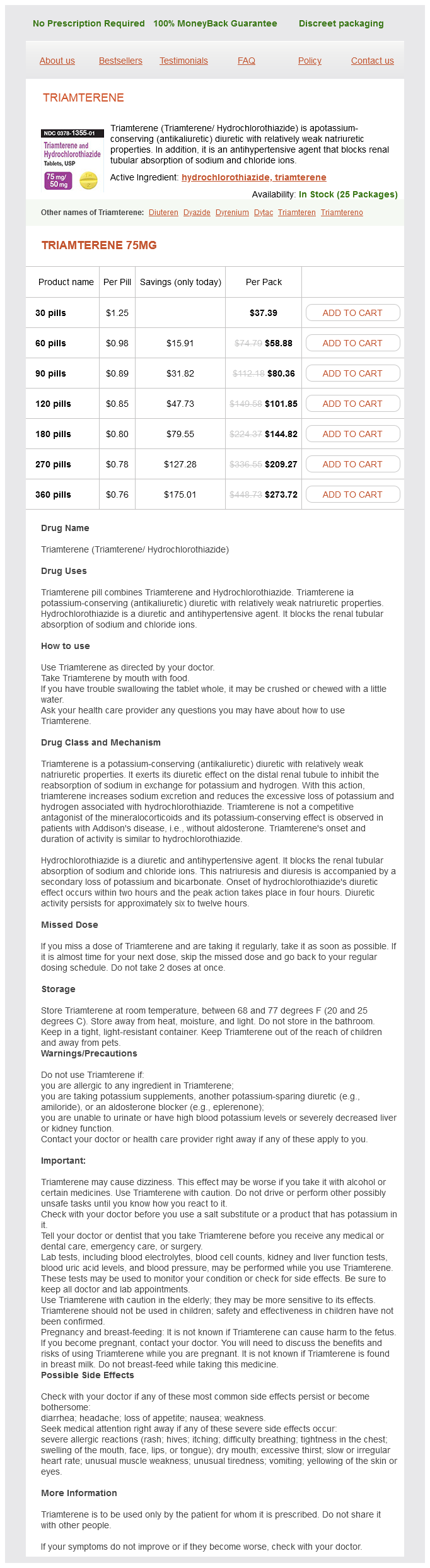
Triamterene Dosage and Price
Triamterene 75mg
- 30 pills - $37.39
- 60 pills - $58.88
- 90 pills - $80.36
- 120 pills - $101.85
- 180 pills - $144.82
- 270 pills - $209.27
- 360 pills - $273.72
Quantitative evaluation of ventricular remodeling can therefore be used to predict clinical outcome and the therapeutic effects of a drug or device intervention arrhythmia reference guide discount triamterene amex. The ensuing ischemia induces an acute injury to the myocardium and initiates an intense inflammatory response. Impaired cardiac systolic and diastolic function and increased filling pressures with pulmonary and systemic venous congestion lead to the syndrome of heart failure. The ventricular remodeling after acute myocardial infarction involves all the different cellular components. The entire myocardial structure needs to be preserved, however, for the cardiomyocytes to exert their contractile function resulting in cardiac systole. The heart is rich in fibroblasts, responsible for creating and maintaining a strong interstitial connective tissue able to support the intense forces associated with cardiac systole. The fibroblasts are also responsible for creating a thick infarct scar opposing the tendency of the infarct area to deform and dilate (aneurysm). Leukocytes are recruited to the heart after ischemic injury, and while inflammation is necessary for infarct healing and debris clearance, an exaggerated inflammatory response can promote further injury and delay infarct healing. The myocardium is composed of cardiomyocytes, endothelial cells, fibroblasts, and resident and infiltrating leukocytes (see also Chapter 4). Reperfusion cytes but accelerates the demise of the nonsalvageable cardiomyocytes (see Chapter 24). Necroptosis also is referred to programmed cell death but while it is an energy-dependent, coordinated process, it shares morphologic features of necrosis. The surviving cardiomyocytes in the border zone display structural and functional changes. These cells often are described as "degenerated," "myofibrillarlytic," "vacuolized," or "autophagic" cardiomyocytes and exist in a delicate balance between death and survival. Increases in the cardiomyocyte crosssectional area or volume (hypertrophy) and development of interstitial fibrosis without a parallel growth in the capillary bed lead to an imbalance between perfusion and demand as a consequence primarily of an impaired diffusion (increased distance between the capillary oxygen content and the cardiomyocyte mitochondria) and an impaired diastolic relaxation and increased intracavitary filling pressures, thereby further reducing the subendocardial perfusion gradient. The imbalance in perfusion-versus-demand and the neurohormonal and proinflammatory milieu promote cardiomyocyte diastolic and systolic dysfunction and transition from prohypertrophy to proapoptosis signaling. Proinflammatory changes in endothelial cells occur within minutes of onset of ischemia and persist for weeks. If the connective tissue was not present or if it had reduced tensile strength, the heart would "break" during systole by "leaking" at the points of least resistance. When the pressure in the heart increases, such as with systemic arterial hypertension or aortic valve stenosis, the collagen content increases in parallel. Leukocyte-derived collagenases further weaken the infarct area by breaking up connective tissue. As a compensatory mechanism to the increased wall stress (stretch), fibroblasts proliferate and differentiate into myofibroblasts, which align along the ventricular wall, providing a partial contraction during systole and producing large amounts of collagen. These changes provide an opposing resistance to the systolic forces that would promote outward excursion of the infarct zone and rupture. Another critical role of the fibroblast is the synthesis of collagen to progressively substitute the infarct area with a fibrotic scar. Endothelial Cells the endothelium regulates the transit of oxygen and nutrients between the blood and the cardiomyocytes. Death of endothelial cells precedes and promotes cardiomyocyte death in ischemia, through the release of proapoptotic factors. There is no restitutio ad integrum (restoration to original condition) after infarction in the heart-largely owing to the fact that once the vascular integrity is compromised, the microvasculature is not reestablished in a functional way. The endothelium also regulates adhesion and migration Leukocytes Leukocytes occupy a central role in the tissue response to injury (see also Chapter 4). Although some degree of inflammation probably is necessary for normal healing, an exuberant inflammatory response promotes myocardial dysfunction, edema, infarct expansion, cell death in cardiomyocytes in the border zone, and worsening systolic and diastolic function. Resolution 453 of the inflammatory response is a physiologic response that limits unnecessary edema, stiffness, and dysfunction. A delay in inflammation resolution, therefore, represents an additional potential pathogenetic mechanism of adverse ventricular remodeling. As such, lack of timely reperfusion due to ineffective treatment 36 or delayed presentation portends an unfavorable prognosis. Impaired tissue-level reperfusion, or "no reflow," relates to persistence of microvascular obstruction despite epicardial coronary artery patency. In the early fibrinolysis trials, the benefits of reperfusion were largest within the first 6 hours and minimal between 13 and 24 hours (see Chapter 15). Prompt reperfusion during acute myocardial infarction prevents adverse ventricular remodeling and heart failure by providing significant, time-dependent myocardial salvage. In the pre-reperfusion era, large transmural infarcts were associated with early cardiac dilation, wall rupture, heart failure, and cardiac death. Impaired tissue reperfusion despite epicardial coronary artery patency ("no reflow") or delayed reperfusion leads to a significantly smaller extent of myocardial damage, with improved clinical outcomes, compared with no reperfusion. Late reperfusion, beyond the window of opportunity for myocardial salvage, is considered to be potentially beneficial in selected patients, providing time- and salvage-independent protection ("open artery hypothesis"). The ratio of the left ventricular diameter to wall thickness reflects the progression of the hypertrophic response relative to the magnitude of dilation. This hypothesis was largely based on longitudinal analysis of data from reperfusion trials in which it was apparent that patients with a patent infarct-related artery had a survival advantage after hospital discharge that remained highly statistically significant even after correction for infarct size and baseline left ventricular ejection fraction. The results of prospective randomized clinical trials, however, only partially support this hypothesis.

ADDII BIOTECH is proud to announce that the company's manufacturing facility located at Baddii, Himachal Pradesh has recently been approved by Food & Drug Authority of Ghana.
Our Manufacturing capability includes a wide range of therapeutic products covering almost every segment.
ADDII BIOTECH given our strong emphasis on product quality and services.